


Can keratitis be detected through a fundoscopic examination?

After developing keratitis, it is essential to select an appropriate treatment method. Corticosteroids may be used for both diagnosis and treatment; however, this approach must be undertaken only under a physician’s guidance. You may also apply a warm towel to the affected eye to promote vasodilation and relieve ocular discomfort—while maintaining strict eye hygiene. Can fundus examination detect keratitis?
Can fundus examination detect keratitis?
Fundus examination alone cannot directly diagnose keratitis. Keratitis is typically assessed using slit-lamp biomicroscopy or direct illumination (e.g., with a penlight). In addition to symptoms such as excessive tearing, photophobia (inability to open the eyes comfortably), various types of ocular discharge, and eye pain, clinicians carefully examine the cornea under illumination for morphological changes—including punctate or patchy epithelial infiltrates, the extent of infiltration, presence of corneal ulcers, corneal transparency, and epithelial surface irregularity or roughness.

Fluorescein staining is also required for accurate assessment. Although the cornea appears transparent to the naked eye under standard illumination, fluorescein sodium staining enhances visualization of epithelial defects and subtle inflammatory changes. Findings may differ between unaided observation and fluorescein staining, as the latter offers greater diagnostic specificity and significantly reduces the risk of missed diagnosis. Once keratitis is confirmed, clinicians determine the likely infectious etiology based on clinical features and perform corneal scraping for microbiological analysis (e.g., Gram stain, culture, PCR) to identify the causative pathogen and guide targeted antimicrobial therapy.
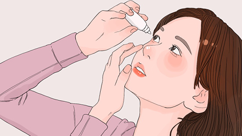
Maintaining proper ocular hygiene is equally critical during treatment. Effective management of keratitis involves not only appropriate pharmacotherapy but also adherence to hygienic practices—such as avoiding eye rubbing, ensuring clean hands before touching the eyes, and properly disinfecting contact lenses or eyewear when applicable. We hope this information is helpful to you.