


Diagnosis of Viral Myocarditis

Patients with viral myocarditis are often overlooked because their prodromal symptoms—such as upper respiratory tract infections—are nonspecific. Therefore, understanding the diagnostic criteria for viral myocarditis is essential for timely intervention. Diagnosis can be confirmed through clinical manifestations, laboratory tests, and auxiliary examinations. So, how is viral myocarditis diagnosed?
Diagnosis of Viral Myocarditis
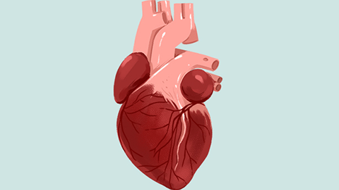
Diagnosis of viral myocarditis relies on electrocardiography (ECG), echocardiography, and cardiac enzyme assays. ECG findings may include ST-segment and T-wave abnormalities, pathological Q waves, and various arrhythmias. Chest X-ray may reveal cardiomegaly (enlarged cardiac silhouette), while echocardiography can demonstrate cardiac dilation—including enlargement of both atria and ventricles—and even intracardiac thrombi. Cardiac magnetic resonance imaging (MRI) may detect focal myocardial signal abnormalities.
Viral myocarditis is an infectious inflammatory disease of the myocardium. During viral epidemics, approximately 5% of infected individuals develop myocarditis, which can be classified as mild, moderate, or severe. Mild cases often present with subtle or nonspecific symptoms. In contrast, severe cases may manifest with fatigue, anorexia, lethargy, palpitations, chest tightness, dyspnea, and pallor. Most patients with viral myocarditis exhibit pallor, fatigue, diaphoresis, and dyspnea.

In daily life, patients are advised to consume nutrient-rich foods—particularly those high in protein and vitamins. We hope this information proves helpful to you.