


Can color Doppler ultrasound check bone density in pregnant women?

In general, color Doppler ultrasound can assess bone density in pregnant women. If necessary, the procedure should be performed under a doctor's guidance. The analysis is as follows:
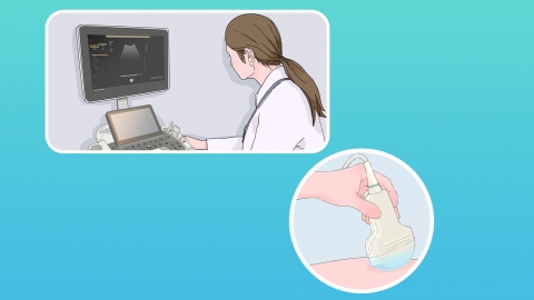
Color Doppler ultrasound, also known as color ultrasound, is a technique that uses ultrasound technology to evaluate bone density. By analyzing the speed of ultrasound conduction and the attenuation of amplitude through bone, it can accurately determine bone content, bone density, and detailed bone structure. This method offers high precision, convenience, and safety, and carries no significant radiation risk, making it highly suitable for bone density assessment in pregnant women.
Additionally, due to hormonal changes and the demands of fetal growth and development during pregnancy, pregnant women are prone to experiencing a decrease in bone density. Bone density testing via color ultrasound can promptly detect calcium reserves in the bones of pregnant women, enabling early identification of declining bone mass trends. This plays a certain role in preventing osteoporosis during pregnancy and ensuring the health of both mother and baby. At the same time, doctors can use the results of color ultrasound to guide pregnant women in appropriately supplementing with nutrients such as calcium and vitamin D, ensuring adequate nutritional support for both mother and child.
Pregnant women should maintain a diverse diet and consume foods rich in minerals such as calcium and phosphorus, such as milk, soy products, and leafy green vegetables.