


Can a 4D color ultrasound detect abnormal kidney development?

Generally, four-dimensional color ultrasound can detect fetal kidney developmental abnormalities, but it has certain limitations. The specific analysis is as follows:
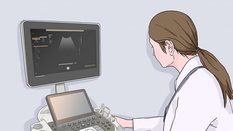
Four-dimensional color ultrasound is an advanced ultrasound examination technique that provides high-resolution images of fetal internal structures, allowing detailed observation of multiple organs including the kidneys. Obvious kidney developmental abnormalities, such as hydronephrosis, polycystic kidney, kidney agenesis, or ectopic kidneys, can usually be accurately identified and diagnosed using four-dimensional color ultrasound.
However, four-dimensional color ultrasound still has certain limitations. For example, it may not accurately detect minor structural or functional abnormalities. Additionally, factors such as fetal position and amniotic fluid volume may sometimes prevent obtaining clear kidney images, thus affecting diagnostic accuracy. If fetal kidney abnormalities are detected via four-dimensional color ultrasound, doctors usually recommend further examinations, such as amniocentesis or chromosomal analysis, to determine the exact cause and nature of the abnormality.
It is recommended to maintain healthy lifestyle habits during pregnancy and undergo regular prenatal checkups to facilitate timely detection and management of any potential issues.