


What can be detected during a mid-pregnancy anomaly scan at 30 weeks of gestation?
Mar 13, 2025
Source: Cainiu Health

Dr. Zhang Lu
Introduction
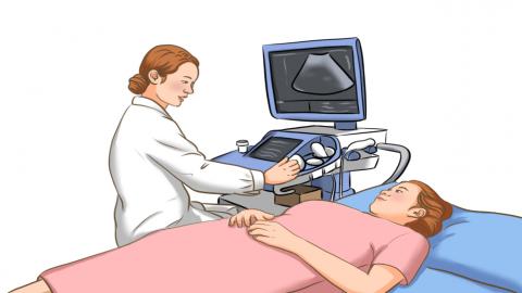
Under normal circumstances, a 30-week prenatal ultrasound examination (minor anomaly scan) can assess various aspects including fetal growth and development, fetal anomaly screening, amniotic fluid analysis, placental condition evaluation, and umbilical cord observation, in order to timely assess the health status of the fetus. If serious fetal diseases or abnormalities are detected through the examination, from the perspective of eugenics, the physician may recommend the pregnant woman to consider termination of pregnancy in a timely manner.
Generally, at 30 weeks of gestation, a minor fetal anomaly scan (minor anomaly screening) can detect conditions related to fetal growth and development assessment, fetal malformation screening, amniotic fluid status analysis, placental condition evaluation, and umbilical cord observation, helping timely assess fetal health. If any abnormalities are detected, timely medical consultation is recommended. Detailed explanations are as follows:
1. Fetal growth and development: Using ultrasound, measurements such as fetal biparietal diameter, head circumference, abdominal circumference, and femur length are taken to assess whether fetal weight and growth align with gestational age. This helps understand fetal growth in utero and identify conditions such as intrauterine growth restriction or macrosomia.
2. Fetal malformations: Minor anomaly screening involves detailed examination of various fetal organ systems, including the cardiovascular, urogenital, digestive, and nervous systems. It can detect certain malformations possibly missed during the major anomaly scan in the second trimester, such as congenital heart disease, limb defects, hydrocephalus, anencephaly, and spina bifida.
3. Amniotic fluid status: Evaluates whether the volume of amniotic fluid is normal, as either excessive or insufficient amniotic fluid may indicate issues in the fetus or mother. For example, polyhydramnios may be associated with fetal malformations, multiple pregnancies, or placental abnormalities; oligohydramnios may be linked to intrauterine growth retardation or placental insufficiency.
4. Placental condition: Examines placental position, maturity, and abnormalities. Abnormal placental position may lead to risks such as placenta previa, which can affect pregnancy outcomes. Premature or delayed placental maturation may also impact fetal nutrition and development.
5. Umbilical cord condition: Assesses whether the umbilical cord is wrapped around the neck and whether umbilical blood flow is normal. Umbilical cord entanglement is a common phenomenon during pregnancy; however, excessive tightness or multiple loops may lead to fetal distress. Abnormal umbilical blood flow may indicate risks such as intrauterine hypoxia.
If severe fetal diseases or abnormalities are detected during examination, doctors may recommend timely termination of pregnancy from the perspective of eugenics. Additionally, minor anomaly scans can provide reference for choosing the mode of delivery—for example, cesarean section may be considered if the fetus is too large, in an abnormal position, or if placental position is abnormal.
References
[1] Li Quanhua, Li Jie, Yang Huixia, et al. Ultrasonographic findings of abdominal pregnancy[J]. Chinese Journal of Medical Imaging Technology, 2025, 41(01): 113-117.
[2] Wu Meimei, Zhong Wanlin. Clinical value of ultrasound screening for fetal malformations at different gestational weeks[J]. Modern Medical Imaging Journal, 2016, 25(05): 922-923.

