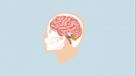
What brain nerve problems can be detected by an EEG?
Electroencephalogram (EEG) can effectively screen for neurological issues such as epilepsy, encephalitis, brain tumors, metabolic encephalopathy, and sequelae of traumatic brain injury by detecting abnormal brain activity. If abnormalities are detected, timely medical consultation is recommended. Detailed analysis is as follows:

1. Epilepsy: EEG can precisely capture abnormal electrical discharges in the cerebral cortex of patients with epilepsy and locate the epileptic focus through multiple leads. Focal seizures may show high-frequency discharges in one brain hemisphere, whereas generalized seizures typically manifest as bilateral synchronized electrical activities, providing key data for epilepsy classification and preoperative evaluation.
2. Encephalitis and Encephalopathy: Viral encephalitis often causes diffuse theta or delta wave activity, with periodic triphasic waves suggesting metabolic encephalopathy. Continuous EEG monitoring allows dynamic observation of disease progression, assessment of the level of consciousness, and guidance in adjusting immunotherapy and dehydration treatments to reduce intracranial pressure.
3. Sequelae of Traumatic Brain Injury: Patients with post-concussion syndrome often show impaired regulation of alpha waves on EEG, while subdural hematomas may present with suppression of beta waves on the affected side. Continuous EEG monitoring helps detect delayed seizures and prevent secondary brain injury.
4. Metabolic Encephalopathy: Hepatic encephalopathy typically presents with bilateral synchronous triphasic waves, while uremic encephalopathy may show widespread slow waves combined with paroxysmal fast rhythms. Combining EEG with laboratory tests allows rapid identification of metabolic causes, guiding treatment strategies such as dialysis or correction of electrolyte imbalances.
5. Brain Tumors: When surrounding brain tissue is compressed or edematous due to a space-occupying lesion, EEG may reveal focal slow waves or spike discharges. Comparing EEG changes before and after surgery helps assess the effectiveness of tumor resection and the functional recovery status of surrounding tissues.
In daily life, maintaining a healthy lifestyle, improving dietary habits, engaging in appropriate physical exercise, and enhancing physical fitness can help reduce the risk of diseases.
[References]
[1] Xie Yanping, Su Qixuan, Li Ping, et al. The Guiding Value of Interictal Scalp EEG in Surgical Decision-Making for Children with Structural Epilepsy[J]. Journal of Modern Electrophysiology, 2025, 32(01): 1-5.
[2] Chen Jiaqi, Xiang Hongying, Ling Yingchun. Study on the Prediction of Cognitive Impairment in Alzheimer's Disease Patients Using EEG Combined with miR-146a-5p[J]. China Modern Doctor, 2025, 63(05): 23-26+56.


