What lesions can be detected by DR imaging?
Mar 19, 2025
Source: Cainiu Health

Dr. Yang Ziqi
Introduction
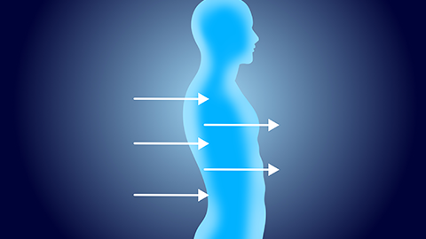
DR examination refers to X-ray examination. In general, X-ray examination can detect lesions of the skeletal system, lung diseases, heart diseases, urinary system abnormalities, gastrointestinal diseases, and other conditions. It is recommended that a qualified physician determine the appropriate imaging examination method based on the patient's specific condition to ensure the most accurate diagnosis and most effective treatment.
DR examination refers to X-ray examination. In general, X-ray examination can detect lesions of the skeletal system, pulmonary diseases, cardiac diseases, urinary system disorders, gastrointestinal diseases, and others. Specific analyses are as follows:
1. Skeletal System Lesions
X-ray examination can clearly demonstrate interruptions in bone continuity, the morphology and orientation of fracture lines, and displacement of fracture ends, enabling the diagnosis of skeletal disorders such as fractures, osteoporosis, arthritis, and bone tumors.
2. Pulmonary Diseases
Conditions such as pneumonia, pulmonary tuberculosis, and lung abscesses can be identified. X-ray examination allows observation of changes in pulmonary markings, uneven density in lung parenchyma, and the presence of abnormal shadows.
3. Cardiac Diseases
X-ray examination can assess the size, shape, and position of the heart, aiding in the diagnosis of cardiac enlargement, pericardial effusion, and other heart diseases. However, for more detailed evaluation of cardiac structure and function, other imaging modalities such as echocardiography or cardiac CT may be required.
4. Urinary System Lesions
X-ray examination can detect high-density stone shadows in the kidneys, ureters, and bladder, assisting in the identification of kidney stones, bladder abnormalities, and ureteral issues.
5. Gastrointestinal Diseases
Conditions such as gastrointestinal obstruction, perforation, space-occupying lesions, ulcers, and inflammation can be diagnosed. X-ray examination is particularly useful in barium meal imaging of the gastrointestinal tract, clearly showing the morphology and pathological changes of the gastrointestinal system.
It is recommended that a qualified physician determine the appropriate imaging method based on the patient's specific condition to ensure the most accurate diagnosis and effective treatment.