


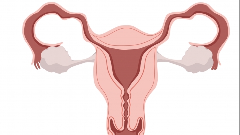
What can be detected by a color Doppler ultrasound of the uterus?

Under normal circumstances, color Doppler ultrasound of the uterus can evaluate the size and shape of the uterus, condition of the endometrium, status of the ovaries, pregnancy-related conditions, and cervical lesions. If discomfort occurs, it is recommended to seek timely diagnosis and treatment at a formal hospital. Detailed analysis is as follows:
1. Uterine Size and Shape
Color Doppler ultrasound uses ultrasound waves penetrating pelvic tissues to generate real-time images, directly measuring the length, width, and thickness of the uterus to determine whether these dimensions fall within the normal range. It can also clearly show whether the uterine shape is regular, helping to identify congenital uterine malformations or morphological abnormalities caused by conditions such as fibroids or adenomyosis.
2. Endometrial Condition
Color Doppler ultrasound can assess the thickness of the endometrium and whether its echogenicity is uniform. If the endometrium appears abnormally thickened or thinned, shows uneven echogenicity, or displays abnormal masses, this may indicate endometrial polyps, hyperplasia, adhesions, or even malignancy. These findings are based on the principle that different tissues reflect ultrasound waves differently, and pathological tissues exhibit imaging characteristics distinct from those of normal endometrium.
3. Ovarian Status
Ultrasound can detect the position and size of both ovaries, observe the number, size, and development of follicles within the ovaries, and identify lesions such as ovarian cysts, polycystic ovary syndrome (PCOS), and ovarian tumors. This is possible because the internal structures of cysts or tumors present characteristic imaging features.
4. Pregnancy-Related Conditions
For pregnant women, color Doppler ultrasound can confirm intrauterine pregnancy by detecting the gestational sac, embryo, and primitive cardiac activity, thereby ruling out ectopic pregnancy. It can also monitor fetal development, placental position, and amniotic fluid depth. These assessments rely on the density differences between embryonic tissues and surrounding tissues, which create clear ultrasound image contrasts.
5. Cervical Lesions
Color Doppler ultrasound can evaluate the thickness and morphology of the cervix, identifying conditions such as cervical hypertrophy, cervical cysts, or cervical fibroids. For suspected lesions within the cervical canal, ultrasound can also assess the extent of the lesion, providing reference information for further diagnostic procedures.
All medical test results must be interpreted in combination with clinical symptoms and other diagnostic tests. If you experience any discomfort or have concerns, it is recommended to seek medical attention promptly and follow the guidance of qualified physicians.