


How is Ankylosing Spondylitis Diagnosed?
Sep 08, 2025
Source: Cainiu Health

Dr. Chen Jian
Introduction
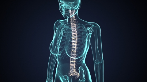
In general, diagnosing ankylosing spondylitis requires a combination of clinical examinations. Common methods include physical examination, blood tests, X-ray examination, CT scan, and magnetic resonance imaging (MRI). Additionally, during the diagnostic process, doctors will comprehensively evaluate symptoms, medical history, and other patient information to ensure accurate and reliable diagnosis.
Generally, a definitive diagnosis of ankylosing spondylitis requires clinical evaluation. Commonly used methods include physical examination, blood tests, X-ray examination, CT scan, and magnetic resonance imaging (MRI). Detailed analysis is as follows:
1. Physical Examination
Doctors assess spinal mobility through specific movements, such as asking the patient to bend forward, extend backward, or laterally flex the lumbar spine, observing whether the range of motion is limited. The sacroiliac joints are also examined by applying pressure or rotating the sacroiliac region to determine if tenderness exists. These assessments can initially detect abnormalities in the spine and sacroiliac joints, providing direction for further diagnosis.
2. Blood Tests
Two key indicators are primarily tested: one is human leukocyte antigen B27 (HLA-B27), which is positive in most patients with ankylosing spondylitis and serves as an important reference; the other involves inflammatory markers such as erythrocyte sedimentation rate (ESR) and C-reactive protein (CRP). Elevated levels of these markers indicate an inflammatory response in the body, aiding in the assessment of disease activity and enhancing diagnostic accuracy when combined with other test results.
3. X-ray Examination
The sacroiliac joints and spine are the primary focus. In early stages, the sacroiliac joints may show blurred or widened joint spaces. As the condition progresses, joint spaces gradually narrow and may even undergo bony fusion. The spine may exhibit a "bamboo spine" appearance, characterized by marginal osteophytes and bridging between vertebrae. This examination clearly reveals structural bone changes.
4. CT Scan
Compared with X-ray examination, CT scans offer higher resolution, allowing clearer visualization of subtle lesions in the sacroiliac joints, such as joint surface erosion and bone sclerosis. CT scans are particularly useful for detecting early lesions or cases where X-ray findings are inconclusive, providing more detailed imaging evidence to aid in accurate diagnosis.
5. Magnetic Resonance Imaging (MRI)
MRI clearly visualizes soft tissues and bone marrow conditions, enabling early detection of lesions such as bone marrow edema and synovitis in the sacroiliac joints, which are difficult to detect using X-ray or CT scans. MRI contributes to early diagnosis, allowing timely intervention and treatment, and also helps assess disease severity.
In addition, during the diagnostic process, doctors also consider the patient's symptoms and medical history to ensure accurate and reliable diagnosis. Once diagnosed, prompt standardized treatment should be initiated to control disease progression.


