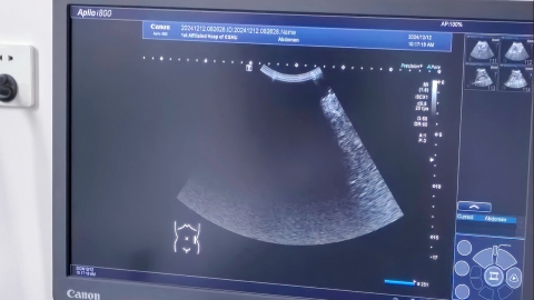
What are the ultrasound findings of gallbladder cancer?
Under normal circumstances, gallbladder cancer exhibits characteristic findings on B-ultrasound examination, primarily involving changes in the gallbladder wall, intraluminal lesions, and surrounding tissues. Common manifestations include gallbladder wall thickening, solid intraluminal masses, gallbladder lumen narrowing or disappearance, gallbladder enlargement or atrophy, and invasion of surrounding tissues. A detailed analysis is as follows:

1. Gallbladder wall thickening: The gallbladder wall shows irregular thickening, often widespread but sometimes localized, with thickness frequently exceeding 3 mm. The thickened wall displays heterogeneous echogenicity, possibly accompanied by areas of increased or decreased echo intensity. The boundary between the thickened wall and normal tissue is often indistinct, and layering disruption or interruption may be observed.
2. Solid intraluminal mass: A solid mass is detected within the gallbladder cavity, typically irregular in shape, poorly demarcated, and with an uneven surface. The mass usually appears hypoechoic or isoechoic with heterogeneous internal echoes. Posterior acoustic enhancement is absent or mild attenuation may be present. The mass is closely attached to and fixed with the gallbladder wall and does not move with changes in body position.
3. Gallbladder lumen narrowing or disappearance: As the tumor grows or the wall thickens, the gallbladder lumen gradually narrows and may completely disappear in severe cases, appearing only as a solid echogenic mass in the gallbladder region. Some patients may have gallstones within the lumen, whose posterior acoustic shadows might obscure the tumor, requiring careful differentiation.
4. Gallbladder enlargement or atrophy: In some early-stage patients, the gallbladder may enlarge with明显 increased volume and wall tension. In advanced stages or with severe tumor progression, the gallbladder may atrophy—becoming smaller in size with markedly thickened walls and loss of contractile function.
5. Invasion of surrounding tissues: As the tumor progresses, it may invade adjacent structures. On ultrasound, the interface between the gallbladder and liver becomes indistinct, and focal hypoechoic areas in the liver suggest hepatic invasion. Enlarged pericholecystic lymph nodes may also be detected, typically round or oval with clear borders and homogeneous or heterogeneous echogenicity, and sometimes showing fusion.
When gallbladder cancer is suspected, further imaging such as CT or MRI is required for definitive diagnosis. Once confirmed, treatment should begin promptly under medical guidance. Maintain a light diet, reduce intake of high-fat and high-cholesterol foods, and support gallbladder health.


