


Can cirrhosis be detected by ultrasound?
Sep 19, 2025
Source: Cainiu Health

Dr. Gao Jun
Introduction
In general, liver cirrhosis can be diagnosed through ultrasound examination, but whether a definitive diagnosis can be made depends on the specific circumstances. In daily life, individuals with high-risk factors for liver disease should undergo regular ultrasound examinations to monitor changes in liver morphology. If abnormalities are detected on ultrasound, timely medical consultation is necessary, and further tests should be completed according to the doctor's recommendations.
In general, liver cirrhosis can be diagnosed through B-ultrasound examination, but whether a definitive diagnosis can be made depends on the specific circumstances. The detailed analysis is as follows:
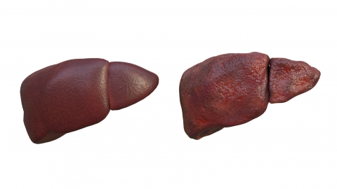
If the patient is in the middle or late stages of liver cirrhosis, obvious changes in liver morphology may already be present—such as reduced liver volume, an uneven liver surface, thickened liver capsule, heterogeneous parenchymal echogenicity, and widened portal vein. When the B-ultrasound images are clear and these characteristic abnormalities are well visualized, and combined with the patient's history of hepatitis or long-term alcohol use, a definitive diagnosis of liver cirrhosis can be supported, thus guiding subsequent treatment.
However, if the patient is in the early stage of liver cirrhosis, significant morphological changes in the liver may not yet be apparent, with only mild alterations in parenchymal echogenicity. In such cases, typical features are difficult to detect via B-ultrasound, and findings may resemble those of chronic hepatitis or fatty liver infiltration. Relying solely on B-ultrasound at this stage makes it challenging to establish a definitive diagnosis; further evaluation using liver function tests, liver fibrosis markers, CT, or MRI is required for a comprehensive assessment.
In daily life, individuals at high risk for liver disease should undergo regular B-ultrasound screenings to monitor changes in liver morphology. If abnormalities are detected on B-ultrasound, prompt medical consultation is recommended, and additional tests should be completed according to physician advice to avoid diagnostic delays. Prior to examination, patients should follow medical instructions for preparation—such as fasting for 8–12 hours—to ensure accuracy of results.
