What are the ultrasound findings of gallstones?
Sep 23, 2025
Source: Cainiu Health

Dr. Gao Jun
Introduction
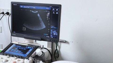
In general, gallstones—solid crystalline substances formed within the gallbladder—appear on B-mode ultrasound as hyperechoic masses with posterior acoustic shadowing, mobility with changes in body position, gallbladder wall alterations, and abnormal bile transmission within the gallbladder. Patients are advised to maintain a low-fat diet, avoid overeating, and minimize stimulation of gallbladder contraction. Regular follow-up gallbladder ultrasounds are recommended to closely monitor the size of the stones.
Under normal circumstances, gallstones—solid crystalline substances formed within the gallbladder—typically present on B-ultrasound as strong echogenic masses, posterior acoustic shadowing, mobility with changes in body position, gallbladder wall alterations, and impaired bile transmission within the gallbladder. A detailed analysis is as follows:
1. Strong echogenic mass: The most typical B-ultrasound finding of gallstones is a strong echogenic mass within the gallbladder. These masses are usually round, oval, or irregular in shape, with clear boundaries. The harder and larger the stone, the more pronounced the echogenicity.
2. Posterior acoustic shadowing: A linear anechoic area often appears behind the strong echogenic mass, known as posterior acoustic shadowing. This results from reflection and absorption of ultrasound waves by the stone. Most gallstones, regardless of size, produce posterior shadowing, which helps differentiate them from other intraluminal gallbladder lesions that typically lack this feature.
3. Mobility with body position: When body position changes, gallstones may shift due to gravity. For example, when moving from a supine to a lateral position, the echogenic mass may move toward the dependent part of the gallbladder. However, if the stone is impacted in the neck of the gallbladder or adhered to the gallbladder wall, it may not move with positional changes.
4. Gallbladder wall changes: Some patients with gallstones also exhibit gallbladder wall thickening. Normally, the gallbladder wall is no thicker than 3 mm; during inflammation, it may increase to over 5 mm. On ultrasound, this appears as uniform or non-uniform wall thickening.
5. Impaired bile transmission within the gallbladder: If gallstones are accompanied by cholecystitis, the ultrasound transmission through bile is reduced, manifesting as fine punctate or flocculent echoes within the bile. This is caused by inflammatory exudates or debris in the bile. Some patients may also develop gallbladder atrophy, characterized by reduced gallbladder volume and decreased bile content.
Patients are advised to maintain a low-fat diet, avoid overeating, and minimize stimulation of gallbladder contraction. Regular follow-up B-ultrasound examinations of the gallbladder are recommended to closely monitor changes in the size and number of stones, and any further interventions should be conducted under medical guidance.