


What are the ultrasound findings of pancreatic cancer?
Sep 23, 2025
Source: Cainiu Health

Dr. Gao Jun
Introduction
In general, pancreatic cancer is a highly malignant tumor of the digestive system. Its ultrasound findings typically include abnormal pancreatic morphology, altered parenchymal echogenicity, dilation of the pancreatic duct, bile duct dilation, and invasion of surrounding pancreatic tissues. Patients are advised to undergo regular follow-up examinations, closely monitor disease progression, and receive treatment under the guidance of qualified physicians.
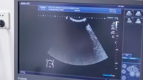
Generally, as a highly malignant tumor of the digestive system, pancreatic cancer typically presents on B-mode ultrasound with abnormalities including altered pancreatic morphology, changes in parenchymal echogenicity, dilation of the pancreatic duct, bile duct dilation, and invasion of surrounding tissues. The specific findings are as follows:
1. Abnormal Pancreatic Morphology: Pancreatic cancer can cause localized enlargement or formation of a mass, leading to distortion of the normal pancreatic shape. In some patients, the pancreatic contour appears unclear and irregular, with indistinct boundaries from surrounding normal tissues, making it difficult to accurately identify the original pancreatic structure.
2. Changes in Parenchymal Echogenicity: Normally, the pancreas exhibits uniform moderate echogenicity on B-ultrasound. In contrast, pancreatic cancer lesions often appear as hypoechoic areas. Some lesions show heterogeneous internal echoes, possibly with punctate or patchy hyperechoic regions. As the disease progresses, the echo characteristics of the lesion may continue to evolve.
3. Pancreatic Duct Dilation: If the tumor compresses or invades the pancreatic duct, it can cause ductal obstruction and subsequent dilation. Ultrasound reveals an increased diameter of the pancreatic duct; in severe cases, a "string-of-beads" appearance may be observed. The dilated duct may extend to the distal pancreas, impairing normal drainage of pancreatic juice.
4. Bile Duct Dilation: When pancreatic cancer is located in the head of the pancreas, it often compresses the common bile duct, causing biliary obstruction and resulting in bile duct dilation. On ultrasound, both intrahepatic and extrahepatic bile ducts appear dilated. Some patients may also present with gallbladder enlargement and thickened gallbladder walls.
5. Invasion of Peripancreatic Tissues: Pancreatic cancer tends to invade adjacent blood vessels and organs. Ultrasound may reveal tumor infiltration of the superior mesenteric vessels, portal vein, etc., leading to vessel wall thickening and luminal narrowing. Additionally, loss of the fat plane around the pancreas may be observed, indicating tumor invasion into surrounding fatty tissues.
Patients are advised to undergo regular follow-up examinations to closely monitor disease progression and receive treatment under the guidance of qualified medical professionals. Maintaining a light diet in daily life, avoiding overeating, and reducing the burden on the pancreas can support recovery.
