What tests can be done to diagnose muscle adhesions?
Sep 25, 2025
Source: Cainiu Health

Dr. Chen Jian
Introduction
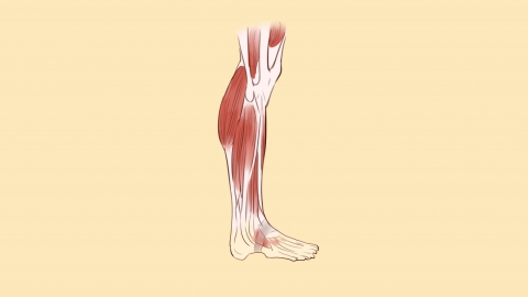
In general, diagnosing muscle adhesions requires combining clinical symptoms with professional examinations. Common diagnostic methods include physical examination, ultrasound, magnetic resonance imaging (MRI), computed tomography (CT), and arthrography. After evaluation, patients should follow professional guidance to develop a rehabilitation or treatment plan based on the diagnosis. It is also important to protect the affected area and avoid excessive activity that could worsen the adhesion.
In general, diagnosing muscle adhesions requires combining clinical symptoms with professional examinations. Common diagnostic methods include physical examination, ultrasound, magnetic resonance imaging (MRI), computed tomography (CT), and arthrography. The details are as follows:
1. Physical Examination: Doctors assess the presence of muscle adhesions through palpation and evaluation of range of motion. During palpation, they can detect muscle tension at the affected site, as well as the presence of nodules or tenderness. Patients may be asked to actively or passively move the relevant joints so that doctors can observe whether their range of motion is restricted.
2. Ultrasound: Ultrasound clearly visualizes the structure of soft tissues such as muscles and tendons. It allows observation of whether muscle fibers are continuous, and whether there are areas of localized thickening or adhesion. It can also determine the extent and severity of adhesions. This method is non-invasive and convenient.
3. Magnetic Resonance Imaging (MRI): MRI provides extremely high soft tissue contrast, enabling detailed visualization of fine structures in muscles, fascia, ligaments, and other tissues. If muscle adhesions are present, MRI can clearly show abnormal signals at the adhesion sites, precisely identifying the location, extent, and relationship of the adhesion to surrounding tissues.
4. CT Scan: Although CT has lower soft tissue resolution compared to MRI, it clearly displays the relationship between muscles and bones. When muscle adhesions are accompanied by changes in bone structure, CT can help determine whether the adhesion affects surrounding bone tissues and rule out similar symptoms caused by bone diseases.
5. Arthrography: For muscle adhesions near joints, arthrography can provide further clarification. During this procedure, contrast dye is injected into the joint cavity, and imaging techniques are used to observe the distribution of the contrast agent. If adhesions are present, the contrast agent will show an abnormal distribution pattern in the affected area.
Prior to undergoing any examination, patients are advised to inform their doctor about the onset time of symptoms, potential triggers, and medical history, so that appropriate tests can be accurately selected. After the examination, patients should follow professional guidance to develop a rehabilitation or treatment plan based on the diagnosis. Additionally, care should be taken to protect the affected area and avoid excessive activity that could worsen the adhesion.