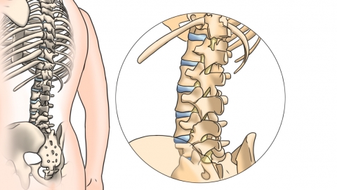
What does a high-density nodule in the thoracic spine mean?
Under normal circumstances, a high-density nodule in the thoracic vertebrae refers to a nodular image with higher density than normal bone or soft tissue observed during imaging examinations of the thoracic spine. Most such nodules are benign, although a small number may carry malignant potential and require further evaluation based on specific test results and clinical symptoms. Detailed analysis is as follows:

A high-density nodule in the thoracic vertebrae may result from osteophyte formation (bone spurs), which can occur due to long-term wear and tear or aging. This leads to bony overgrowths forming nodular high-density images. These are typically asymptomatic and do not require special treatment. Daily management includes avoiding excessive physical strain and performing appropriate back and core muscle exercises. Alternatively, the nodule may represent callus formation remaining after healing of a thoracic vertebral fracture. During fracture healing, accumulated bone callus may form a nodular appearance; if asymptomatic, regular follow-up imaging to monitor changes is sufficient.
In rare cases, a high-density thoracic nodule may be associated with benign conditions such as enostosis (bone island) or osteochondroma. A bone island is a dense focus of compact bone within spongy bone, while osteochondroma is a common benign bone tumor. These conditions generally do not require treatment but should be monitored periodically through imaging to assess any changes in size or shape. If the nodule is accompanied by symptoms such as pain or restricted movement, further diagnostic evaluation is necessary to determine its nature.
In daily life, it is important to maintain good posture, avoid prolonged bending or sitting, and reduce stress on the thoracic spine. Regular imaging follow-ups are recommended to monitor the nodule's progression.


