


How to differentiate thyroid nodules from thyroid cancer
Nov 05, 2025
Source: Cainiu Health

Dr. Yang Ziqi
Introduction
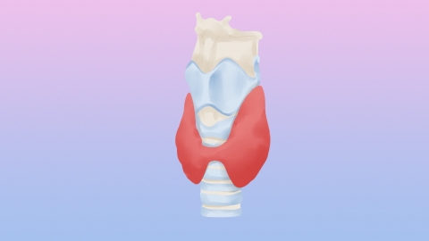
In general, thyroid nodules are mostly benign, while thyroid cancer is a malignant tumor. Differentiating between the two requires a comprehensive evaluation including ultrasound examination, nodule characteristics, growth rate, laboratory tests, and pathological analysis. Key points for differentiation include ultrasound imaging features, nodule texture and margins, differences in growth rate, thyroid function markers, and results of pathological examination.
Under normal circumstances, thyroid nodules are mostly benign, while thyroid cancer is a malignant tumor. Differentiating between the two requires comprehensive evaluation including ultrasound imaging, nodule characteristics, growth rate, laboratory tests, and pathological examination. Key points for differentiation include ultrasound features, nodule texture and margins, differences in growth speed, thyroid function markers, and pathological findings. A detailed analysis is as follows:
1. Ultrasound Imaging Features: Benign nodules typically appear regular in shape—round or oval—with uniform internal echogenicity, no calcification or only coarse calcifications, and minimal blood flow signals. In contrast, thyroid cancer nodules often have irregular shapes, poorly defined borders, heterogeneous echogenicity, microcalcifications, and markedly increased (abnormal) blood flow signals.
2. Nodule Texture and Margins: On physical examination, benign nodules tend to be soft or of moderate firmness, with clear boundaries and good mobility, without adhesion to surrounding tissues. Malignant thyroid nodules are usually hard, have indistinct borders, poor mobility, and may feel fixed due to adhesion to adjacent structures. They are often non-tender upon palpation.
3. Growth Rate Differences: Benign nodules grow slowly and generally do not affect surrounding tissues. Thyroid cancer nodules grow more rapidly, with noticeable increases in size over a short period. As they enlarge, they may compress nearby structures, leading to symptoms such as difficulty swallowing or voice changes.
4. Thyroid Function Markers: Blood tests assessing thyroid function and related antibodies can aid in differentiation. Most patients with benign nodules have normal thyroid function and antibody levels within the normal range. Some patients with thyroid cancer may present with abnormal thyroid function, and certain rare subtypes may show specific tumor markers.
5. Pathological Examination Results: Histopathological analysis is the gold standard for distinguishing benign from malignant nodules. Fine-needle aspiration cytology (FNAC) involves extracting a small number of cells from the nodule for microscopic evaluation. The presence of cancer cells confirms thyroid cancer, whereas benign cellular features support a diagnosis of a benign nodule.
If a thyroid nodule is detected, patients should undergo regular follow-up examinations to monitor changes in the nodule and follow professional medical advice regarding appropriate diagnostic and treatment strategies to ensure optimal thyroid health.


