


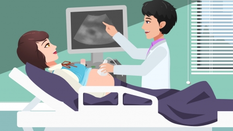
What should I do if I’ve had more than ten 4D ultrasounds without success?

Failure to obtain satisfactory 4D ultrasound images after multiple attempts (e.g., 10–15 times) is commonly attributable to suboptimal fetal positioning, excessive maternal abdominal fat thickness, oligohydramnios (low amniotic fluid volume), nuchal cord (umbilical cord around the fetal neck), or fetal atrioventricular septal defect. Appropriate adjustments based on individual circumstances—or targeted interventions—can help facilitate successful re-examination. Detailed explanations follow:
1. Suboptimal Fetal Positioning
When the fetus remains in a fixed position for an extended period, key anatomical structures—including the face, limbs, and heart—may be obscured, preventing acquisition of complete imaging data. To encourage fetal repositioning, mothers may engage in gentle physical activity such as slow walking or climbing stairs; lightly massage the abdomen; or consume a small amount of sweet food to stimulate fetal movement.
2. Excessive Maternal Abdominal Fat Thickness
Thick abdominal adipose tissue attenuates ultrasound signal transmission, resulting in blurred images and reduced clarity in visualizing fetal anatomy. Prior to the examination, moderate physical activity may help minimize abdominal fat laxity and accumulation. During scanning, alternating between supine and lateral decubitus positions may improve image quality.
3. Oligohydramnios (Low Amniotic Fluid Volume)
Insufficient amniotic fluid reduces the cushioning space surrounding the fetus, restricting fetal mobility and significantly impairing ultrasound image resolution. Under medical guidance, mothers may take Yiqi Weixue Capsules, Baoling Yunbao Oral Solution, or Vitamin C effervescent tablets. Daily intake of warm water and consumption of fresh fruits and vegetables are also recommended to maintain adequate hydration.
4. Nuchal Cord (Umbilical Cord Around the Fetal Neck)
A nuchal cord limits fetal mobility, hindering comprehensive multi-angle imaging. As directed by a healthcare provider, mothers may take calcium gluconate oral solution, ferrous lactate oral solution, and vitamin B complex tablets. Resting in the left lateral decubitus position daily and closely monitoring fetal movements are advised.
5. Fetal Atrioventricular Septal Defect (AVSD)
This congenital cardiac anomaly involves abnormal development of the atrioventricular septum, resulting in structural gaps that compromise precise assessment of cardiac anatomy via 4D ultrasound. Prenatal serial follow-up monitoring is essential. After birth, mild cases may be managed medically with digoxin tablets, spironolactone tablets, and hydrochlorothiazide tablets; severe defects typically require surgical repair—atrioventricular septal defect correction.
Prior to undergoing 4D ultrasound during pregnancy, women should proactively optimize their physical condition: maintaining regular sleep-wake cycles, adhering to a balanced diet, engaging in appropriate physical activity according to individual tolerance, completing scheduled antenatal examinations, and promptly identifying and addressing any abnormalities—to safeguard normal fetal development.