


What precautions should be taken for a patellar bone crack?

For a patellar fissure (crack in the kneecap), attention should be paid to immobilization and protection of the affected limb, elevation of the limb to reduce swelling, appropriate physical therapy, standardized medication use, and regular follow-up monitoring. A patellar fissure is a type of bone crack injury; premature weight-bearing may cause displacement of the crack, impairing bone healing. If severe knee swelling and pain, purplish discoloration or numbness of the skin occur, seek prompt medical evaluation and treatment.
1. Immobilization and Protection of the Affected Limb: Immobilization and protection are the top priorities. Use a brace or plaster cast to stabilize the injured knee joint and avoid flexion, extension, or twisting movements. Do not bear weight or walk too early, as this may displace the fracture site. Minimize movement of the affected limb in daily life to protect the damaged bone and create a stable local environment conducive to bone repair.
2. Elevation of the Affected Limb to Reduce Swelling: During rest, elevate the injured leg above heart level using soft padding. This promotes venous and lymphatic return from the lower extremity, thereby reducing knee swelling and hematoma. When lying supine, place a soft cushion under the leg; avoid prolonged dependency of the limb, which may exacerbate swelling and alleviate local distension and discomfort.
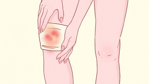
3. Appropriate Physical Therapy: Physical therapy should be tailored to the stage of injury. Apply cold compresses within the first 48 hours post-injury to constrict blood vessels and minimize exudation. After 48 hours, switch to warm compresses to accelerate absorption of hematoma. Carefully control temperature and duration during therapy; avoid aggressive rubbing, massaging, or pressing on the affected area to prevent secondary injury to the fractured site.
4. Standardized Medication Use: Follow professional guidance for medication use. Topical medications should be applied to the skin surrounding the knee to relieve localized pain and swelling. For significant pain, oral analgesics may be used as adjunctive therapy to alleviate discomfort. Monitor for any skin reactions or systemic side effects during treatment; do not adjust dosage without medical advice. Combine pharmacotherapy with adequate rest to optimize recovery.
5. Regular Follow-up Monitoring: Schedule timely imaging examinations (e.g., X-ray or MRI) to assess actual progress of bone healing. Adjust immobilization duration and rehabilitation plans based on clinical recovery. If healing is delayed or suboptimal, promptly modify management strategies to prevent complications such as delayed union or fracture displacement.
During convalescence, maintain a diet rich in protein and calcium. Avoid unguided knee flexion and extension exercises. Ensure adequate warmth for the lower limb and gradually resume activity in a stepwise manner to prevent functional deterioration of the knee joint.


