


How to Determine Whether It Is Cholangiocarcinoma

Cholangiocarcinoma (bile duct cancer) can be diagnosed through a comprehensive evaluation encompassing clinical symptoms, blood tests, imaging screening, tumor marker assays, and histopathological biopsy. If you experience rapid jaundice (yellowing of the skin and sclera), persistent abdominal pain accompanied by hematemesis (vomiting blood), or other concerning symptoms, prompt medical consultation and treatment are strongly advised.
1. Clinical Symptoms: As the disease progresses, patients may develop jaundice, dull upper abdominal pain, and decreased appetite. The skin and sclera gradually turn yellow; urine darkens while stools become pale. Some individuals experience unexplained weight loss. While these symptoms provide valuable preliminary clues, they alone cannot confirm a diagnosis.
2. Blood Tests: Venous blood samples are analyzed to assess liver function parameters—including bilirubin and alkaline phosphatase—both of which typically rise in cholangiocarcinoma due to biliary obstruction and associated hepatic injury. Inflammatory markers may also be evaluated. However, blood tests alone cannot definitively determine the nature of the lesion.
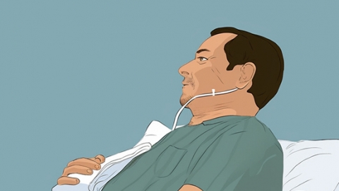
3. Imaging Screening: Ultrasound, computed tomography (CT), or magnetic resonance imaging (MRI) are employed to visualize bile duct morphology—assessing ductal dilation, wall thickening, and presence of space-occupying lesions. These modalities precisely localize the lesion and evaluate the extent of involvement in surrounding tissues, making them widely used first-line screening tools in clinical practice.
4. Tumor Marker Assays: Serum tumor markers (e.g., CA19-9, CEA) are measured via blood testing. Elevated levels may occur in cholangiocarcinoma but are not specific: benign biliary conditions such as inflammation or stones can also cause similar elevations. Therefore, results must always be interpreted in conjunction with other diagnostic findings.
5. Histopathological Biopsy: A small sample of suspicious tissue is obtained—via endoscopic retrograde cholangiopancreatography (ERCP), percutaneous transhepatic cholangiography (PTC), or surgical resection—and examined microscopically for cellular atypia and architectural abnormalities. This remains the gold standard for definitive diagnosis, enabling accurate differentiation between benign and malignant lesions and guiding subsequent therapeutic planning.
In daily life, maintain a light, low-fat diet and minimize intake of high-fat foods. Ensure adequate rest and strictly avoid tobacco and alcohol. Should any unusual physical changes arise, do not delay seeking medical attention; undergo comprehensive diagnostic evaluations promptly to clarify the nature and extent of biliary tract pathology.


