


What are the specific symptoms of melasma?

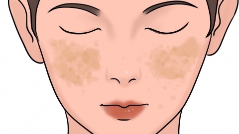
Chloasma (melasma) manifests as light brown macules on the face, with indistinct borders, variable pigmentation intensity, symmetrical distribution, and no significant pruritus or pain. These hyperpigmented patches commonly appear on the cheeks, forehead, and perioral area. Exposure to sunlight and hormonal fluctuations can progressively worsen the condition. If the pigmented lesions rapidly enlarge over a short period or are accompanied by localized erythema, swelling, or ulceration, prompt medical evaluation and treatment are recommended.
1. Light brown facial macules:
In the early stage, lesions typically appear as faint brown patches that gradually darken as the condition progresses. The patches are flat—neither raised above nor depressed below the skin surface—and feel identical to surrounding normal skin upon palpation. Melanin accumulates in the epidermis, resulting in visible pigmentary changes.
2. Indistinct borders:
The margins of the patches lack sharp definition and blend gradually into adjacent normal skin, without forming a distinct, ring-like demarcation. Some patches may slowly extend outward, merging with neighboring areas of hyperpigmentation, thereby continuously increasing the overall affected area.
3. Variable pigmentation intensity:
Pigment intensity often intensifies noticeably following sun exposure, sleep deprivation, or hormonal fluctuations. Conversely, with adequate sun protection and rest, the discoloration may slightly fade. Pigment activity fluctuates with overall physiological status, leading to periods of relative improvement and exacerbation.
4. Symmetrical distribution:
Hyperpigmented lesions most commonly appear bilaterally on the cheeks, with corresponding locations on the left and right sides of the face. The forehead, nasal bridge, and perioral region may also be involved. This symmetrical pattern is associated with systemic influences such as endocrine changes and ultraviolet radiation exposure.
5. Absence of significant pruritus or pain:
The affected areas generally do not cause itching or stinging sensations; the epidermal surface remains intact, with no associated papules or vesicles. In some individuals, generalized facial dryness may occur, but any discomfort is usually attributable to impaired skin barrier function rather than the pigmented lesions themselves.
Maintain regular sleep-wake cycles, manage emotional stress, minimize prolonged direct sun exposure, adopt gentle and simplified skincare routines, and consistently support skin barrier health to help prevent progressive worsening of chloasma.


