What Is Bone Age Assessment?
May 16, 2022
Source: Cainiu Health

Dr. Chen Jian
Introduction
Bone age—also known as skeletal age—is determined by analyzing specific features visible on X-ray images of bones. Bone age assessment primarily relies on characteristic changes in bone morphology and structure across different age groups. For instance, during childhood and adolescence, bones progressively mature with advancing age, including epiphyseal closure. These specific skeletal landmarks are observed to estimate bone age.
Bone age assessment determines an individual’s biological age by evaluating skeletal development. The analysis is as follows:
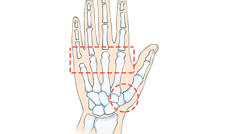
Bone age—also termed skeletal age—is determined using characteristic radiographic features of bones visible on X-ray images. Bone age assessment primarily relies on age-related morphological and structural changes in the skeleton. For instance, during childhood and adolescence, bones progressively mature with advancing chronological age, including epiphyseal closure. By observing these specific skeletal landmarks, bone age can be estimated with reasonable accuracy.
Wrist X-ray radiography is the most commonly used method for bone age assessment. By obtaining X-ray images of the wrist, clinicians can evaluate skeletal development in this region—particularly the degree of epiphyseal plate closure—to determine bone age. Alternatively, bone age may be assessed using laser-based bone densitometry: scanning the tibia to evaluate both bone mineral density (BMD) and bone age. Since BMD changes closely correlate with skeletal growth and maturation, this technique also serves as a valid tool for bone age estimation.
Bone age reflects an individual’s biological age, which may differ from their chronological age. In children and adolescents, bone age helps predict potential height gain and plays a valuable role in monitoring growth and development, as well as facilitating early detection of growth abnormalities.
If bone age assessment is required, individuals should visit a reputable hospital and undergo the procedure under the guidance of qualified physicians following standardized protocols, to ensure safety, accuracy, and reliability of results. Prior to the examination, patients should remain relaxed and avoid excessive tension or anxiety, as stress-induced muscle tension may interfere with accurate assessment of skeletal status.