


Typical Symptoms and Treatment of Molluscum Contagiosum

Molluscum contagiosum is a common skin disease with contagious potential; thus, early detection and prompt treatment are essential upon diagnosis.
Typical Symptoms and Treatment of Molluscum Contagiosum
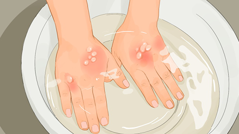
The characteristic cutaneous manifestation of molluscum contagiosum includes dome-shaped, soft, translucent papules exhibiting a waxy luster. Lesions typically feature central umbilication, from which a white, cheese-like molluscum body can be expressed. Primary treatment is local. The molluscum bodies may be completely removed using forceps or a curette, followed by topical application of medicated ointment. Caustic topical agents may also be applied directly. Prophylactic measures are crucial—maintain good personal hygiene and avoid sharing bathtubs, swimming pools, or other communal facilities irregularly. Upon confirmed diagnosis, patients should be promptly isolated and treated until clinical cure is achieved.
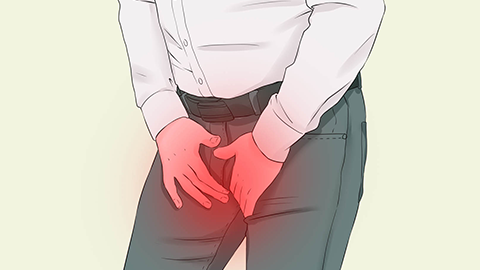
Molluscum contagiosum is a frequently encountered infectious dermatosis in both daily life and clinical practice. It is classified as a nationally notifiable infectious disease in China; therefore, immediate isolation and timely reporting to public health authorities are mandatory following diagnosis. It predominantly affects adolescents and children, with lesions commonly appearing on the neck, trunk, lower abdomen, and external genitalia.
Molluscum contagiosum is primarily caused by direct contact with the molluscum contagiosum virus (MCV). Diagnosis relies on integration of clinical history, characteristic lesion morphology, and supportive laboratory tests when indicated. We hope this information proves helpful!