


How is alveolar osteitis treated?

Alveolar osteitis—commonly known as “dry socket”—is clinically managed according to the following principles: debridement, isolation from external stimuli, and promotion of granulation tissue formation.Regarding etiology, the most widely accepted theory is the fibrinolytic theory: trauma or infection triggers fibrinolysis, leading to disintegration of the blood clot, kinin formation, and consequently severe pain.
How is alveolar osteitis treated?
The first step in treatment is local debridement—irrigating the socket with hydrogen peroxide and normal saline. The second step involves either gently curetting the socket using specialized instruments to induce bleeding, followed by placement of a cotton pellet to protect the newly formed clot; or inserting an iodoform gauze strip into the socket to isolate it from external stimuli and create optimal conditions for healing. Both approaches typically result in healing within 7–10 days. Clinical experience suggests that curettage-induced bleeding yields superior outcomes.
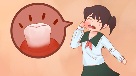
Clinically, dry socket manifests as osteitis of the bony walls of the extraction socket—or, in milder cases, as localized osteomyelitis. Based on the presence or absence of tissue necrosis, it is classified as either necrotic (or “septic”) or non-necrotic (“non-septic”); the necrotic type is far more common in clinical practice. Patients typically develop persistent pain 3–4 days after tooth extraction, often radiating to the cheek. Clinical examination reveals surrounding gingival erythema and swelling, obvious necrotic debris within the socket, an empty-looking alveolus, foul odor, and marked tenderness upon palpation of the bony socket walls.
Dry socket exhibits a distinct predilection: it occurs predominantly in mandibular molars. Among the three molars, the third molar (i.e., the wisdom tooth) has the highest incidence, followed by the first molar; other teeth are rarely affected.
We hope the above information is helpful to you.