


What fetal surface abnormalities can be detected by 4D color ultrasound?
Mar 05, 2025
Source: Cainiu Health

Dr. Zhang Lu
Introduction
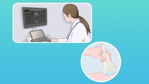
Under normal circumstances, four-dimensional color ultrasound can detect fetal surface abnormalities such as abnormalities in the head, face, limbs, cardiac structure, and genitalia. Although four-dimensional color ultrasound can provide detailed information about the fetus, it cannot rule out all potential issues. Pregnant women are advised to undergo regular prenatal examinations to monitor fetal development.
Generally, four-dimensional color ultrasound can detect fetal surface abnormalities such as abnormalities of the head, face, limbs, cardiac structure, and genitalia. Detailed analysis is as follows:
1. Head Abnormalities
Four-dimensional color ultrasound utilizes the principle of ultrasound reflection. When ultrasound waves penetrate fetal head tissues, different tissue interfaces produce varying intensities of reflected echoes. These echoes are captured by the device and converted into images, clearly displaying the structure of the fetal brain, such as the ventricles and cerebral parenchyma.
2. Facial Abnormalities
Ultrasound waves encountering different tissues of the fetal face generate varying reflection signals due to differences in acoustic impedance. These signals are collected and used to create high-resolution three-dimensional images, visually displaying the morphology and positional relationships of facial organs such as the lips, nose, eye sockets, and ears.
3. Limb Abnormalities
Ultrasound beams can scan fetal limbs segment by segment, clearly showing structures such as the limbs and trunk, helping to identify any malformations, such as limb deficiencies, spina bifida, abnormal spinal shape, skeletal developmental anomalies, and hand or foot deformities.
4. Cardiac Structural Abnormalities
Ultrasound waves reflecting from different layers of cardiac tissue and valves can clearly display structures such as the four cardiac chambers, ventricular septum, atrial septum, and heart valves.
5. Genital Abnormalities
Ultrasound can display the tissue morphology and structure of the fetal genital area, allowing observation of the appearance and structure of the fetal genitalia to determine whether there are developmental abnormalities such as hypospadias, cryptorchidism, or labial fusion.
Pregnant women are advised to undergo regular prenatal checkups to promptly monitor their own health status and fetal development.
