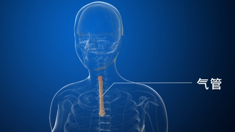
What are the causes of tracheal deviation to the affected side?
Generally, tracheal deviation to the affected side may be caused by external trauma, postural changes, pneumothorax, pleural effusion, atelectasis, and other reasons. Patients should seek timely medical evaluation at a regular hospital to determine the cause and receive appropriate treatment. Detailed explanations are as follows:
1. External Trauma
If the tracheal area suffers external impact, it may lead to temporary displacement of the trachea. This condition is usually accompanied by symptoms such as pain and swelling, but generally does not cause long-term effects. It can be relieved through physical therapies like cold or hot compresses, and adequate rest with avoidance of strenuous physical activity is recommended.
2. Postural Changes
In certain positions, such as prolonged lateral decubitus positioning, slight tracheal deviation may occur. This is considered a normal phenomenon and requires no special treatment. Adjusting body position, maintaining proper sitting and sleeping postures, and avoiding prolonged immobility are recommended.

3. Pneumothorax
During pneumothorax, air enters the pleural cavity, causing compression of the lung tissue on the affected side and increased intrathoracic pressure, which pushes the trachea toward the healthy side. Patients often experience sudden chest pain and dyspnea, and severe cases may present with subcutaneous emphysema. Diagnosis can be confirmed via chest X-ray or CT scan. Small pneumothoraces may be managed with observation and oxygen therapy, while large pneumothoraces require chest tube insertion according to medical advice.
4. Pleural Effusion
Large-volume pleural effusion increases pressure on the affected side of the thoracic cavity, compressing lung tissue and displacing mediastinal structures. Common causes include tuberculous pleuritis and pleural metastasis of malignant tumors. Symptoms generally include cough, chest tightness, and manifestations of the underlying disease.
5. Atelectasis
In cases of atelectasis, the affected lobe or segment of the lung collapses due to the inability of air to enter, reducing the lung's supportive effect on the thoracic cavity on that side and causing the trachea to shift toward the affected side. Accompanying symptoms may include dyspnea, chest pain, and cough. If necessary, patients can receive oxygen therapy or surgical treatment at a regular hospital.
When tracheal displacement is detected, timely imaging studies should be conducted to determine the underlying cause, and strenuous activity should be avoided to prevent worsening of dyspnea. Maintaining airway patency in daily life is important, and patients with chronic lung disease should quit smoking and prevent infections. Immediate medical attention is required if acute symptoms such as marked stridor or orthopnea occur.


