How to Determine Cardiovascular Blockage
Jul 21, 2025
Source: Cainiu Health

Dr. Tian Hongbo
Introduction
Cardiovascular blockage often presents with chest pain, typically a pressing pain behind the sternum that may radiate to the left shoulder and back, accompanied by symptoms such as chest tightness, shortness of breath, and sweating. Some symptoms worsen during physical exertion or emotional excitement and may be relieved by rest. An electrocardiogram (ECG) can record the heart's electrical activity and may show characteristic changes such as ST-segment alterations and abnormal T waves, especially when the symptoms occur.
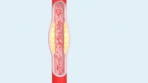
Generally, the assessment of cardiovascular blockage involves observing symptom presentation, performing electrocardiogram (ECG) testing, conducting blood tests, implementing imaging examinations, and undertaking angiography. Detailed explanations are as follows:
1. Observing symptom presentation: Cardiovascular blockage often presents with chest pain, typically a pressing discomfort behind the breastbone that may radiate to the left shoulder and back, accompanied by chest tightness, shortness of breath, and sweating. Symptoms may worsen with exertion or emotional excitement and may improve with rest.
2. Performing electrocardiogram (ECG) testing: An ECG records the heart's electrical activity. Characteristic changes such as ST-segment elevation or T-wave abnormalities may appear during cardiovascular blockage. Testing during symptom onset can provide initial evidence for identifying blockage.
3. Conducting blood tests: Measuring biomarkers of myocardial injury, such as troponin and creatine kinase-MB (CK-MB), can aid in assessing the severity of blockage. These markers rise when myocardial ischemia and necrosis occur due to cardiovascular blockage.
4. Implementing imaging examinations: Echocardiography allows evaluation of cardiac structure and function. Localized abnormalities in myocardial motion may indicate vascular blockage in the corresponding region. Chest CT can visualize the morphology of large vessels such as the aorta and help identify potential blockages.
5. Undertaking angiography: Angiography is a precise method for diagnosing cardiovascular blockage. By injecting contrast agent into the blood vessels, imaging equipment can clearly visualize the course and lumen condition of the vessels, accurately identifying the location, extent, and severity of the blockage.
If symptoms suggestive of cardiovascular blockage occur, physical activity should be stopped immediately and rest is advised. Prompt medical evaluation is essential. Once diagnosed, treatment should follow medical advice. Additionally, adopting healthy lifestyle habits such as a low-salt, low-fat diet and regular exercise is important, along with controlling risk factors to reduce the likelihood of worsening blockage.