What tests are needed for patients with hydatidiform mole?
Jul 22, 2025
Source: Cainiu Health

Dr. Zhang Lu
Introduction
In general, hydatidiform mole is a gestational trophoblastic disease. Patients need to undergo examinations such as B-ultrasound, serum human chorionic gonadotropin (hCG) testing, fetal heart rate monitoring, X-ray examination, and histological examination. Before undergoing these examinations, patients should inform their doctors about their medical history, symptoms, and other relevant information, so that doctors can better formulate treatment plans.
Generally, hydatidiform mole is a gestational trophoblastic disease. Patients need to undergo B-ultrasound examination, serum human chorionic gonadotropin (hCG) testing, fetal heart monitoring, X-ray examination, histological examination, and other related tests. Detailed analysis is as follows:
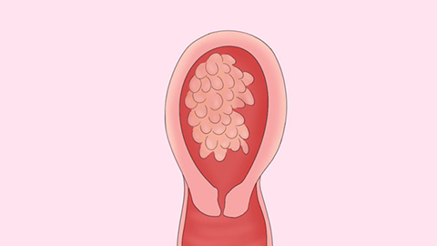
1. B-Ultrasound Examination
B-ultrasound examination is one of the important auxiliary diagnostic methods for hydatidiform mole. Using a high-frequency probe, the grape-like structures within the uterine cavity, as well as cystic structures and fluid accumulation within the placenta, can be clearly observed.
2. Serum Human Chorionic Gonadotropin (hCG) Testing
Serum human chorionic gonadotropin testing is an important indicator for diagnosing hydatidiform mole. Patients with hydatidiform mole typically have serum hCG levels higher than those of normal pregnancy, and these levels continue to rise after 8 to 10 weeks of gestation. This helps in diagnosing and differentiating hydatidiform mole from gestational trophoblastic tumors.
3. Fetal Heart Monitoring
Fetal heart monitoring can help determine whether the fetal heartbeat is normal. In normal pregnancies, Doppler ultrasound can detect fetal heart sounds after two months, but in cases of hydatidiform mole, only uterine blood flow noises can be heard.
4. Chest X-Ray Examination
Hydatidiform mole may metastasize to the lungs. A chest X-ray helps detect any lung metastases early, which is helpful in assessing disease severity and formulating a treatment plan. If suspicious shadows are found in the lungs, the location, size, and shape of the lesion can be identified under a physician's guidance, enabling targeted treatment.
5. Histological Examination
Histological examination is the definitive method for diagnosing hydatidiform mole. During a curettage procedure, all removed tissues must be sent for histological analysis. By observing cellular morphology and structure, hydatidiform mole can be confirmed, and its characteristics can be determined.
Prior to undergoing these examinations, patients should inform their physician about their medical history and symptoms to assist in developing an effective treatment plan. Additionally, patients should maintain a positive mindset and actively cooperate with their physician's treatment and management recommendations.