Can B-mode ultrasound detect liver cirrhosis?
Jun 07, 2022
Source: Cainiu Health

Dr. Jin Zhongkui
Introduction
B-mode ultrasound (B-ultrasound) is one of the diagnostic tests for liver cirrhosis and can generally detect this condition. Imaging techniques such as B-ultrasound and CT are used to assess the liver’s status—including its size, surface smoothness, presence of nodules, and thickening of the portal vein—providing key evidence for physicians to diagnose liver cirrhosis. Viral infection and chronic alcohol intoxication are the primary causes of liver cirrhosis.
Most of the time, you may need to visit a hospital for examination due to bodily pain or abnormalities in certain areas. Among various diagnostic tests, B-mode ultrasound (B-ultrasound) not only provides clearer visualization of internal body structures but also plays a valuable role in disease detection. So, can B-ultrasound detect liver cirrhosis?
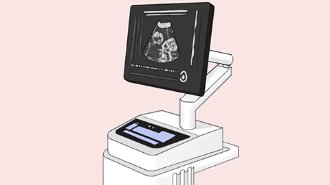
Can B-ultrasound detect liver cirrhosis?
B-ultrasound is one of the standard imaging examinations used in diagnosing liver cirrhosis and is generally capable of detecting it. By employing imaging techniques such as B-ultrasound and CT scans, physicians assess the liver’s condition—including its size, surface smoothness, presence of nodules, and thickening of the portal vein—these findings collectively serve as key diagnostic criteria for liver cirrhosis. B-ultrasound can indeed identify cirrhosis, with characteristic features clearly visible on the image, notably an undulating or “wavy” surface contour. Additionally, B-ultrasound may reveal associated signs such as liver atrophy, splenomegaly, or portal vein dilation. However, if a patient exhibits only highly suspicious clinical features of cirrhosis, B-ultrasound alone cannot definitively confirm the diagnosis; further diagnostic testing is therefore required.
Patients with liver cirrhosis should cultivate healthy lifestyle habits, maintain a positive mental outlook, and ensure balanced nutrition. Moreover, they must avoid indiscriminate use of medications and undergo regular follow-up examinations to better control their condition. Liver protection is essential—providing nutritional support to damaged hepatocytes promotes their recovery and helps preserve normal liver function.

Patients need not be overly anxious; by paying attention to daily lifestyle factors, disease progression can be effectively managed. We hope this information has been helpful to you.