What Are the Differences Between Hyperplasia and Tumors?
Mar 13, 2022
Source: Cainiu Health

Dr. Qi Zhirong
Introduction
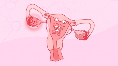
Hyperplasia refers to an increase in the number of cells within a tissue or organ, often resulting in enlargement of the tissue or organ. Hyperplasia can be physiological or pathological. A tumor is an abnormal growth (neoplasm) arising from localized cellular proliferation within the body, characterized by abnormal cell morphology, growth patterns, and function. Tumors are classified as either benign or malignant.
Hyperplasia and tumors may differ in definition, pathogenesis, pathological features, diagnosis, and treatment. It is recommended to consult a healthcare provider to determine the underlying cause and follow medical advice for appropriate management. The following analysis elaborates on these differences:
1. Definition
Hyperplasia refers to an increase in the number of cells within a tissue or organ, often resulting in enlargement of the affected tissue or organ. It can be physiological—such as uterine hyperplasia during pregnancy to accommodate fetal growth—or pathological, caused by factors including hormonal imbalance, chronic irritation, or genetic mutations. A tumor, by contrast, is an abnormal growth (neoplasm) arising from localized, uncontrolled cell proliferation within the body. Tumors exhibit abnormal cellular morphology, growth patterns, and function. They are classified as either benign or malignant: benign tumors grow slowly, have well-defined borders, and do not metastasize; malignant tumors grow rapidly, display poorly defined margins, and readily invade surrounding tissues and metastasize to distant sites.
2. Pathogenesis
Hyperplasia typically arises in response to physiological demands or pathological stimuli. Physiological hyperplasia represents a normal adaptive response to bodily needs, whereas pathological hyperplasia may result from multiple factors—including hormonal imbalances, chronic irritation, or genetic mutations. Tumor development involves complex interactions among various factors, such as genetic mutations, dysregulation of the cell cycle, and aberrant intracellular signaling pathways. These disruptions collectively lead to uncontrolled cellular proliferation and neoplastic formation.
3. Pathological Features
In hyperplasia, cellular morphology and function remain largely normal, and the process is usually reversible. Although structural changes may occur in the affected tissue or organ, functional impairment is generally minimal. In contrast, tumor cells exhibit marked abnormalities in both morphology and function, with invasive and metastatic potential. While benign tumor cells closely resemble their normal counterparts in appearance and function, malignant tumor cells show significant deviations. Malignant cells can infiltrate adjacent tissues and disseminate via hematogenous or lymphatic routes to distant organs.
4. Diagnosis
Diagnosis of hyperplasia commonly relies on histopathological examination and imaging studies. Histopathology helps determine the nature and extent of hyperplasia, while imaging enables assessment of its morphology and anatomical location. Similarly, tumor diagnosis requires integration of histopathology, imaging modalities, and tumor marker assays. Histopathology establishes tumor type and stage; imaging evaluates tumor size, location, and morphology; and tumor marker testing aids in characterizing tumor biology and predicting prognosis.
5. Treatment
Physiological hyperplasia typically requires no intervention. Pathological hyperplasia necessitates targeted therapy addressing the underlying cause—for example, eliminating irritants or correcting hormonal imbalances. Severe cases may warrant surgical excision. Benign tumors are usually managed effectively with complete surgical resection and carry an excellent prognosis. Malignant tumors demand multimodal therapy—including surgery, chemotherapy, radiotherapy, and/or targeted therapy—tailored to individual patient needs. Prognosis for malignancy depends on tumor type, stage at diagnosis, and appropriateness of therapeutic strategy.
If a tumor is diagnosed, prompt treatment is strongly advised. During treatment, maintaining a positive emotional state and avoiding excessive emotional stress is beneficial. Additionally, patients should adhere to regular rest schedules and avoid overexertion.