How is anoscopy performed?
Aug 24, 2022
Source: Cainiu Health

Dr. Cheng Yicheng
Introduction
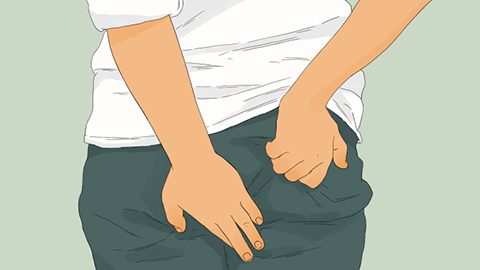
During anoscopy, the knee-chest position is typically adopted. Prior to anoscopy, visual inspection of the anus is performed to assess for any abnormalities in its shape and to check for the presence of abscesses. A rubber glove is worn on the examining finger, which is then lubricated with liquid paraffin. First, the examiner gently inserts a finger into the anal orifice to allow the patient to acclimatize, followed by digital rectal examination.
Modern medical technology has advanced significantly, and diagnostic equipment has been further refined. An anoscope is a 7-cm-long instrument whose internal diameter varies, enabling direct visualization of lesions in the anal canal and distal rectum. So, how is an anoscopy performed?
How Is Anoscopy Performed?
During anoscopy, the patient typically assumes the knee-chest position. Prior to insertion, the anus is visually inspected for abnormalities in shape or signs of abscesses. A rubber glove is worn on the examining finger, which is then lubricated with liquid paraffin. First, the examiner gently inserts a finger into the anal opening to allow the patient to gradually acclimate; this is followed by digital rectal examination to assess for anal or rectal stenosis, inflammation, tenderness, or other abnormalities.
Anoscopy is the optimal method for diagnosing anorectal disorders and also serves as an important therapeutic tool. Several types exist—including circular (round) and oblique (slanted) designs. The standard circular anoscope facilitates unilateral injection or ligation of hemorrhoids. Another type—the two-blade split anoscope—is primarily used for minor anorectal surgical procedures or anal canal dilation, and is generally not employed for routine diagnostic examination.

In daily life, patients should maintain a positive mindset, actively cooperate with their physicians during treatment, take prescribed medications regularly and as directed, and pay attention to routine self-care—thereby promoting prompt clinical improvement. We hope this information is helpful to you.