


How is a fallopian tube imaging procedure performed?
Dec 27, 2024
Source: Cainiu Health

Dr. Zhang Lu
Introduction
Under normal circumstances, the main steps of hysterosalpingography include preoperative preparation, adjusting body position, local disinfection, exposing the cervix, injecting contrast medium, and observing the results. If any discomfort occurs, it is recommended to seek timely medical attention at a hospital and undergo standardized treatment under a physician's guidance. During the procedure, the examinee should stay as relaxed as possible to avoid fallopian tube spasms caused by pain or tension.
Generally, the main steps of hysterosalpingography include preoperative preparation, adjustment of body position, local disinfection, exposure of the cervix, injection of contrast medium, and observation of results. If discomfort occurs, it is recommended to seek timely medical consultation and follow standardized treatment under a doctor's guidance. A detailed explanation is as follows:
1. Preoperative Preparation
The procedure should be scheduled between 3 to 7 days after the end of menstruation, as this period carries a lower risk of genital tract infection and the endometrium is thinner, reducing the likelihood of endometriosis. Before the procedure, the bladder should be emptied. Patients with constipation may require an enema to maintain the normal position of the uterus. An iodine allergy test must be completed beforehand, and only individuals with negative test results can undergo the imaging procedure. If allergic to iodinated oil, patients should not undergo iodinated oil imaging. Thirty minutes before the procedure, patients should receive an intramuscular injection of atropine sulfate as directed by a physician to relieve spasms and promote relaxation.
2. Adjustment of Body Position
The patient should assume either the lithotomy or supine position, with legs apart to expose the perineum, facilitating the physician's operation.
3. Local Disinfection
The physician will disinfect the instruments and the patient's external genitalia and vaginal areas to prevent infection.
4. Exposure of the Cervix
The physician will use a speculum to dilate the vagina, fully exposing the cervix to facilitate subsequent procedures. The vaginal fornix and cervix will be disinfected again.
5. Injection of Contrast Medium
The physician will insert a catheter into the cervical opening and slowly inject the contrast medium into the uterine cavity and fallopian tubes. The contrast medium should fill the catheter and uterine cavity completely, and air should be expelled.
6. Observation of Results
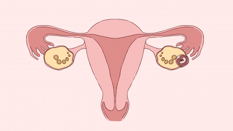
With the assistance of X-ray imaging, the physician observes the area under examination, assessing the visualization of the contrast medium within the fallopian tubes and pelvic cavity. This helps determine whether the fallopian tubes are patent or obstructed, identifies the location of any blockage, and evaluates the morphology of the uterine cavity.
During the imaging procedure, the patient should remain as relaxed as possible to avoid fallopian tube spasms caused by pain or anxiety, which could falsely suggest tubal occlusion.
