


What are transvaginal ultrasound and hysterosalpingography?
Nov 15, 2025
Source: Cainiu Health

Dr. Zhang Lu
Introduction
Transvaginal ultrasound and hysterosalpingography are two commonly used gynecological and reproductive system examinations. Transvaginal ultrasound uses ultrasound imaging to observe pelvic organs, while hysterosalpingography employs contrast agents to visualize and assess the patency of the fallopian tubes. These two examinations serve different purposes, and their application should be determined based on specific medical conditions. Before choosing an examination, it's important to clarify individual needs—for example, women experiencing prolonged infertility during pregnancy planning may prioritize hysterosalpingography.
Transvaginal ultrasound and hysterosalpingography are two commonly used gynecological and reproductive system examinations. Transvaginal ultrasound uses ultrasound imaging to observe pelvic organs, while hysterosalpingography employs contrast agents to assess the patency of the reproductive tract. These two tests serve different purposes, and the choice between them should be based on specific medical conditions. The detailed analysis is as follows:
Transvaginal ultrasound, short for transvaginal sonography, involves inserting an ultrasound probe into the vagina. It does not require a full bladder and can clearly display the morphology and structure of pelvic organs such as the uterus, ovaries, and fallopian tubes. This examination is non-invasive and easy to perform. It can be used to monitor follicular development and diagnose conditions such as uterine fibroids and ovarian cysts. It is especially suitable for women who are trying to conceive or in early pregnancy, allowing earlier detection of pregnancy-related conditions.
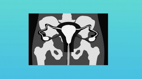
Hysterosalpingography, commonly referred to as tubal imaging, involves injecting a contrast agent into the uterine cavity and fallopian tubes via a catheter, followed by X-ray or ultrasound imaging to observe the flow of the contrast medium. This helps determine whether the fallopian tubes are open, whether there are blockages, and the location of any obstructions. The procedure should be performed 3–7 days after menstruation ends. It is minimally invasive, and some individuals may experience temporary bloating or abdominal pain. Rest and hygiene should be maintained after the examination.
Before choosing an examination method, it's important to clarify individual needs. For example, women experiencing prolonged infertility may prioritize hysterosalpingography, whereas routine gynecological checkups or ovulation monitoring are better suited to transvaginal ultrasound.