


What heart problems can an electrocardiogram (ECG) detect?
Mar 17, 2025
Source: Cainiu Health

Dr. Tian Hongbo
Introduction
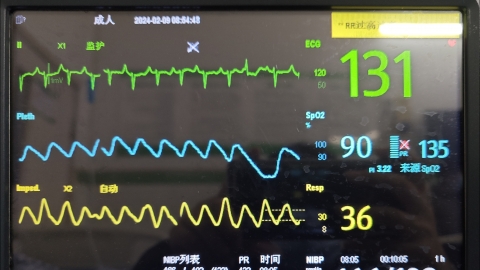
Under normal circumstances, electrocardiogram (ECG) is a common non-invasive diagnostic test used to evaluate the electrical activity of the heart. It can detect various cardiac issues, primarily including arrhythmias, myocardial ischemia, myocardial infarction, cardiac hypertrophy, and electrolyte imbalances. ECG is one of the important tools in diagnosing heart diseases and provides significant evidence for the diagnosis of various cardiac conditions.
Generally, electrocardiogram (ECG) is a common non-invasive diagnostic test used to evaluate cardiac electrical activity. It can detect various cardiac problems, mainly including arrhythmias, myocardial ischemia, myocardial infarction, cardiac hypertrophy, and electrolyte disturbances. Detailed analysis is as follows:
1. Arrhythmias
An ECG can clearly detect abnormalities in cardiac rhythm, including sinus tachycardia or bradycardia, atrial premature beats, atrial fibrillation, etc. These abnormal rhythms may manifest as abnormally fast or slow heartbeats, along with changes in the morphology and rhythm of P waves and QRS complexes.
2. Myocardial Ischemia
When there is insufficient blood supply to the myocardium, the ECG may show ST-segment depression, flattened or inverted T waves. During angina attacks in patients with coronary artery disease, the ECG may display ST-T changes in corresponding leads, indicating myocardial ischemia.
3. Myocardial Infarction
Myocardial infarction occurs when coronary artery occlusion leads to ischemic necrosis of the myocardium. The ECG shows typical changes during different phases of myocardial infarction. In the acute phase, the ECG typically presents with ST-segment elevation in a convex upward pattern, presence of abnormal Q waves, and tall T waves.
4. Cardiac Hypertrophy
The ECG can help identify structural abnormalities of the heart. Left ventricular hypertrophy manifests on ECG as increased QRS complex voltage, which may be accompanied by ST-T changes. Poorly controlled hypertension can lead to left ventricular hypertrophy, presenting as left ventricular high voltage on ECG.
5. Electrolyte Disturbances
Imbalances in electrolytes such as potassium and calcium can affect the electrophysiological activity of the myocardium, producing characteristic changes on ECG. In hypokalemia, the ECG may show flattened or inverted T waves, prominent U waves, and prolonged QT interval. Severe hypokalemia may lead to ventricular arrhythmias.
The ECG is one of the important tools for diagnosing heart diseases and provides essential evidence for the diagnosis of various cardiac conditions. However, it has limitations, as some heart diseases may not leave obvious traces on the ECG. Therefore, physicians usually make a comprehensive judgment by combining the patient's symptoms, physical signs, and results from other diagnostic tests.
References:
[1] Wang Xin. "Evaluation of the value of electrocardiogram combined with echocardiography in assessing cardiac function in patients with myocardial infarction." Modern Medical Imaging Journal, 2024, 33(12): 2317-2320.
[2] Jiang Xiaoqing. "The role of lead aVR in electrocardiographic diagnosis of ventricular hypertrophy." Gansu Medical Journal, 2018, 37(05): 448-449.


