Can uterine fibroids be detected by B-ultrasound?
Jul 09, 2025
Source: Cainiu Health

Dr. Zhang Lu
Introduction
In general, uterine fibroids can be detected through B-ultrasound. Before undergoing a B-ultrasound examination, patients receiving an abdominal scan should have a moderately full bladder to obtain clearer images; patients undergoing a transvaginal scan do not need to retain urine but should avoid the menstrual period. The results must be interpreted by a physician in conjunction with clinical symptoms. For smaller fibroids, regular follow-up B-ultrasounds may be necessary to monitor changes.
Under normal circumstances, uterine fibroids can be detected through B-ultrasound. Detailed analysis is as follows:
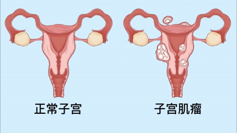
Uterine fibroids are solid masses formed by the proliferation of uterine smooth muscle cells. Their density differs from that of the surrounding normal uterine muscle tissue. B-ultrasound can generate clear images through ultrasound reflection, accurately revealing information such as the size, number, location of the fibroid, and its relationship with the uterine muscle layer. Both transabdominal and transvaginal ultrasounds can effectively detect uterine fibroids, especially those larger than 1 cm in diameter, making it a commonly used and effective method for clinical screening and diagnosis.
Before undergoing a B-ultrasound examination, patients receiving transabdominal ultrasound need to moderately fill their bladder by holding urine to obtain clearer images; transvaginal ultrasound does not require bladder filling but should be avoided during menstruation. The results must be interpreted comprehensively by a physician in conjunction with clinical symptoms. Smaller fibroids may require regular follow-up ultrasounds to monitor changes. If ultrasound suggests the presence of uterine fibroids, further evaluation by a healthcare professional is necessary to determine whether treatment is required, avoiding both undue anxiety and neglect of the condition.