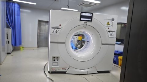
How many regions are included in a whole abdominal CT scan?
Generally, a full abdominal CT scan usually covers multiple anatomical regions, mainly including the upper abdomen, middle abdomen, lower abdomen, pelvis, peritoneum, and mesentery. The specific analysis is as follows:

1. Upper Abdomen: The upper abdomen contains organs such as the liver, gallbladder, spleen, stomach, duodenum, and pancreas. A full abdominal CT scan can clearly display the morphology, size, and position of these organs, helping to identify any abnormalities such as space-occupying lesions, inflammation, or stones.
2. Middle Abdomen: The middle abdomen mainly includes the small intestine, colon, kidneys, and upper ureters. CT examination can visualize the structures of these organs and help determine whether there are conditions such as intestinal obstruction, kidney hydronephrosis, or stones.
3. Lower Abdomen: The lower abdomen includes the colon, lower ureters, and bladder. A full abdominal CT scan can detect abnormalities in these organs, such as intestinal tumors or bladder stones.
4. Pelvis: The pelvis is an important part of a full abdominal CT scan. In males, the pelvic region includes the prostate and seminal vesicles; in females, it includes the uterus, fallopian tubes, and ovaries. The scan can detect abnormalities such as tumors, inflammation, or fluid accumulation within the pelvis.
5. Peritoneum and Mesentery: The peritoneum lines the inner surfaces of the abdominal and pelvic cavities, while the mesentery connects the intestines and contains blood vessels and lymph nodes. A full abdominal CT scan can reveal whether the peritoneum is thickened or whether there is lymph node enlargement or masses in the mesentery, aiding in the assessment of conditions such as peritonitis or tumor metastasis.
Before undergoing a full abdominal CT scan, it is important to follow preparation instructions, such as fasting and retaining urine, to ensure image clarity. After the examination, drinking plenty of water helps eliminate the contrast agent. Maintaining a healthy lifestyle can also help reduce the risk of abdominal organ diseases.


