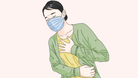
What causes atelectasis?
Generally, atelectasis may be caused by factors such as shallow breathing, airway secretion blockage, bacterial pneumonia, pulmonary tuberculosis, or benign lung tumors. It is recommended to seek timely medical consultation to determine the exact cause and receive appropriate treatment under a physician's guidance. Detailed analysis is as follows:

1. Shallow breathing: Patients who are bedridden for long periods or recovering post-surgery often experience shallow and slow breathing, insufficient lung expansion, and reduced air volume in the alveoli, which can lead to atelectasis. This commonly occurs in the lower lobes of both lungs and is accompanied by chest tightness, shortness of breath that worsens with physical activity. Regular assistance with turning over and sitting up, guidance on deep breathing exercises, daily balloon-blowing exercises to improve lung function, and promoting lung expansion can help reduce the occurrence of atelectasis.
2. Airway secretion blockage: Secretions such as phlegm and mucus can block the bronchi, preventing airflow from reaching the corresponding lung tissues and causing atelectasis. This may be accompanied by coughing and difficulty expectorating, with diminished local breath sounds upon auscultation. Drinking plenty of water helps thin secretions. Follow medical advice to use ambroxol oral solution or acetylcysteine effervescent tablets to promote expectoration. When necessary, nebulization inhalation can be used to humidify the airways, aiding secretion removal and relieving obstruction.
3. Bacterial pneumonia: Bacterial infections, such as those caused by Streptococcus pneumoniae, can trigger inflammation, leading to increased secretions that block the airway and cause atelectasis. Symptoms may include high fever, purulent sputum, chest pain, and blood tests indicating bacterial infection. Patients should rest in bed and follow medical instructions for antibiotic treatment, such as intravenous penicillin sodium, cefotaxime sodium injection, or levofloxacin sodium chloride injection. Additionally, postural drainage can help expel sputum and restore lung ventilation.
4. Pulmonary tuberculosis: Infection with Mycobacterium tuberculosis damages lung tissue, forming caseous necrotic material that blocks the bronchi, leading to atelectasis. Symptoms may include low-grade fever, night sweats, hemoptysis, and a positive tuberculin test. Patients must strictly follow medical advice for anti-tuberculosis treatment, such as combination therapy with isoniazid tablets, rifampicin capsules, and pyrazinamide tablets. The treatment course must be completed in full. Regular chest CT scans are necessary to monitor recovery from atelectasis, and discontinuation of medication without medical consultation should be avoided.
5. Benign lung tumors: Tumor growth can block the bronchi, obstruct airflow, and cause atelectasis, accompanied by coughing and chest tightness, with symptoms worsening as the tumor enlarges. Imaging studies may reveal space-occupying lesions. Patients should promptly determine the tumor's nature and follow medical recommendations for thoracoscopic tumor resection to remove the tumor and relieve bronchial obstruction, restoring lung tissue ventilation. Regular follow-up after surgery is necessary to prevent recurrence.
In daily life, smoking should be avoided, and exposure to irritant gases such as dust and smoke should be minimized to reduce lung tissue damage. For long-term bedridden patients, enhanced nursing care is essential, including regular changes in body position and encouragement of effective coughing and expectoration. Engaging in appropriate aerobic exercise can strengthen lung function and respiratory muscles, reducing the risk of atelectasis.


