


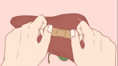
How is a hemangioma on the liver treated?
Aug 21, 2025
Source: Cainiu Health

Dr. Gao Jun
Introduction
In general, hepatic hemangiomas may be caused by genetic factors, hormonal fluctuations, abnormal proliferation of vascular endothelial cells, intrahepatic vascular malformations, abnormal repair following liver injury, and other factors. It is recommended to seek timely medical attention, determine the size and location of the hemangioma, and then follow the doctor's guidance for improvement through regular monitoring, medication, surgical treatment, or other appropriate methods.
Under normal circumstances, liver hemangiomas may be caused by genetic factors, hormonal fluctuations, abnormal proliferation of vascular endothelial cells, intrahepatic vascular malformations, abnormal repair following liver injury, and other factors. It is recommended to seek timely medical consultation. After clarifying the size and location, improvement can be achieved under a doctor's guidance through regular monitoring, medication, surgical treatment, and other approaches. A detailed analysis is as follows:
1. Genetic factors: A family history of liver hemangiomas may predispose individuals to abnormal vascular development within the liver due to genetic influences. If the hemangioma's diameter is less than 5 cm and no discomfort is present, a liver ultrasound should be reviewed every 6-12 months to monitor size changes. Alcohol consumption should be avoided in daily life to reduce liver irritation.
2. Hormonal fluctuations: Elevated estrogen levels during female puberty or hormonal changes during pregnancy may stimulate the growth of hepatic blood vessels, leading to hemangioma formation. Avoid taking estrogen-containing supplements, maintain a regular lifestyle, and reduce hormonal imbalances. If there is no significant enlargement of the hemangioma, its growth typically ceases once hormone levels stabilize.
3. Abnormal proliferation of vascular endothelial cells: Excessive proliferation of vascular endothelial cells within the liver may form cavernous hemangiomas, which are often asymptomatic when small. If the hemangioma's diameter is between 5-10 cm, medications such as propranolol tablets, prednisolone tablets, or methylprednisolone tablets may be used under medical guidance to inhibit cell proliferation.
4. Intrahepatic vascular malformations: Abnormal differentiation of liver blood vessels during embryonic development may result in congenital hemangiomas, which may gradually enlarge in adulthood. If the hemangioma's diameter is less than 10 cm and there are no compressive symptoms, regular monitoring should continue; if dull pain appears in the upper right abdomen, transarterial embolization may be performed to block the blood supply to the hemangioma, preventing further growth.
5. Abnormal repair after liver injury: Following liver trauma from impact or surgery, abnormal proliferation during the vascular repair process may lead to hemangioma formation, potentially accompanied by discomfort in the liver area. If the hemangioma's diameter exceeds 10 cm or compresses surrounding tissues, surgical removal of the liver hemangioma should be performed to completely excise the affected tissue and relieve the compression.
In daily life, strenuous exercise should be avoided to prevent abdominal trauma that could damage the hemangioma. Maintain a regular lifestyle and avoid late nights, which can increase the liver's metabolic burden. If symptoms such as severe upper right abdominal pain or bloating occur, timely follow-up is necessary to adjust the treatment plan and maintain liver health.


