


How to examine neurofibromatosis
Aug 21, 2025
Source: Cainiu Health

Dr. Yang Ziqi
Introduction
In general, neurofibromas can be examined through methods such as physical examination, ultrasound, magnetic resonance imaging (MRI), pathological biopsy, and genetic testing. Additionally, computed tomography (CT) scans may also be used for neurofibroma evaluation. It is also important to maintain a positive mindset and actively cooperate with the doctor throughout the examination process, both before and after the tests.
Generally, neurofibromas can be diagnosed through physical examination, ultrasound, magnetic resonance imaging (MRI), pathological biopsy, genetic testing, and other methods. Detailed explanations are as follows:
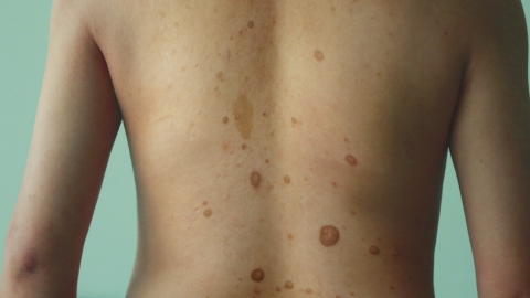
1. Physical Examination
Doctors can initially assess the presence of symptoms related to neurofibromas by observing whether café-au-lait spots or subcutaneous nodules exist on the skin, palpating the texture and mobility of nodules, and checking for abnormal neurological signs. During the examination, patients should cooperate with the doctor to expose the entire body's skin, especially hidden areas.
2. Ultrasound Examination
Using ultrasound waves to scan subcutaneous and deep tissues can clearly display the tumor's location, size, shape, internal structure, and its relationship with surrounding tissues, helping detect neurofibromas on the body surface and within internal organs. Patients should prepare according to the doctor's instructions before the examination—for example, fasting may be required for abdominal scans.
3. Magnetic Resonance Imaging (MRI)
Detailed internal body images are generated using magnetic fields and radiofrequency pulses. MRI can accurately show the relationship between neurofibromas and surrounding tissues such as nerves, blood vessels, and bones, clarifying the tumor's extent of invasion and providing important evidence for diagnosis and treatment planning. Patients must remove all metal objects and remain still during the examination.
4. Pathological Biopsy
A small tissue sample is taken from the affected area and analyzed microscopically and pathologically to determine the tumor's nature and type. This is considered the gold standard for diagnosing neurofibromas. Wounds should be kept clean after the biopsy to prevent infection.
5. Genetic Testing
By analyzing blood or tissue samples, mutations in related genes such as NF1 and NF2 can be detected. This helps diagnose hereditary neurofibromas, determine disease type, and assess genetic risk. No special preparation is needed before testing; sample collection should follow the doctor's instructions.
In addition, computed tomography (CT) scans and other methods might also be used during neurofibroma examinations. It is important to maintain a positive mindset and actively cooperate with the doctor throughout the diagnostic process.


