What tests are needed for ankylosing spondylitis?
Generally, the diagnosis of ankylosing spondylitis requires a combination of imaging, laboratory indicators, and clinical evaluation. Commonly used tests include imaging of the sacroiliac joints, HLA-B27 genetic testing, inflammatory marker testing, physical examination of the spine and joints, and examination of peripheral joints and eyes. The specific analyses are as follows:

1. Imaging of the Sacroiliac Joints: This is a key test for diagnosing the condition. Common imaging methods include X-ray, CT, and magnetic resonance imaging (MRI). X-rays can initially reveal whether there is bone sclerosis, joint space narrowing, or fusion, making them suitable for initial screening. CT provides higher resolution and can clearly show early bone erosions and blurred joint surfaces.
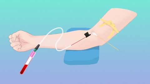
2. HLA-B27 Genetic Testing: This test is performed via blood sampling. The HLA-B27 gene is strongly associated with ankylosing spondylitis, with approximately 90% of patients testing positive. While a positive result alone cannot definitively diagnose the disease, it can enhance diagnostic accuracy when combined with symptoms and imaging findings. A negative result does not completely rule out the disease and must be interpreted in conjunction with other tests.
3. Inflammatory Marker Testing: This primarily includes erythrocyte sedimentation rate (ESR) and C-reactive protein (CRP), both measured through blood sampling. Elevated levels indicate inflammatory activity in the body, aiding in diagnosis, assessing disease severity, and providing reference for adjusting treatment plans.
4. Physical Examination of the Spine and Joints: Doctors evaluate spinal and joint mobility through specific movements, such as asking the patient to bend forward, turn around, or look upward, to assess lumbar flexion, lateral bending, and cervical range of motion. Palpation of the sacroiliac joints helps determine the presence of tenderness and joint involvement.
5. Examination of Peripheral Joints and Eyes: Some patients may have involvement of peripheral joints. Doctors check for joint swelling, tenderness, and assess joint mobility. Eye examination is also necessary to observe for conjunctival congestion, photophobia, or other symptoms, to identify extra-articular manifestations such as uveitis, providing comprehensive information for disease evaluation.
After completing the examinations, doctors will make a comprehensive assessment based on the results to confirm or exclude the diagnosis. Patients with confirmed diagnosis should follow medical advice for treatment, undergo regular follow-up tests, monitor disease progression, promptly adjust treatment plans, and maintain joint function and quality of life.


