


How to calculate the size of the gestational sac
Nov 14, 2025
Source: Cainiu Health

Dr. Zhang Lu
Introduction
To calculate gestational sac size, methods generally include ultrasound measurement of diameters, using the mean diameter formula, estimating in conjunction with gestational age, referring to standard reference tables, and dynamic monitoring with serial comparisons. Ultrasound measurement of diameters: The length, width, and height of the gestational sac are measured via B-mode ultrasound. The clearest cross-sectional view of the sac is selected during measurement to ensure accuracy. Measurements are typically recorded in millimeters.
To calculate the size of the gestational sac, methods generally include ultrasound measurement of diameters, using the mean diameter formula, estimating in conjunction with gestational age, referring to standard value tables, and dynamic monitoring with comparative analysis. Specific details are as follows:
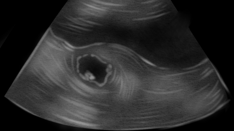
1. **Ultrasound measurement of diameters**: Use B-mode ultrasound to measure the length, width, and height (three diameters) of the gestational sac. The clearest cross-sectional image of the sac should be selected during measurement to ensure accuracy. Measurements are typically recorded in millimeters and serve as the foundational data for calculating gestational sac size; subsequent calculations or assessments require formulas or reference standards.
2. **Using the mean diameter formula**: Add the measured values of length, width, and height, then divide the sum by 3 to obtain the mean diameter, which represents the overall size of the gestational sac. This method reduces errors from single-diameter measurements and more objectively reflects the actual volume of the sac. It is a commonly used, simple calculation approach in clinical practice.
3. **Estimation based on gestational age**: Different gestational ages correspond to relatively fixed ranges of gestational sac sizes. For example, at around 6 weeks of gestation, the mean diameter is approximately 15 mm, and about 25 mm at 7 weeks. By knowing the gestational age, one can roughly estimate the expected size of the sac and determine whether it matches the actual developmental status.
4. **Referring to standard value tables**: In medicine, standardized tables for gestational sac size have been established based on extensive statistical data. These tables specify the normal ranges of length, width, height, and mean diameter corresponding to each gestational week. Comparing measured values with these standard ranges allows for rapid assessment of whether the sac's development falls within the normal interval.
5. **Dynamic monitoring and comparison**: Repeat the ultrasound examination after an interval of 1–2 weeks, measure the sac size again, and compare it with previous results to observe the growth rate. Under normal conditions, the gestational sac increases in size according to a predictable pattern each week. Slowed or stalled growth may indicate abnormal development and warrants further evaluation.
If gestational sac size needs to be assessed, it is recommended to undergo ultrasound examination at a qualified medical facility, where trained professionals can perform accurate measurements and interpret the results. If discrepancies between sac size and gestational age or abnormal growth patterns are detected, follow the physician’s advice to complete further evaluations, identify underlying causes, and initiate timely interventions as needed.

