


Can Onychomycosis Heal Spontaneously?
Aug 06, 2022
Source: Cainiu Health

Dr. Liu Wan
Introduction
Onychomycosis is a fungal infection. Factors that may contribute to its development include nail trauma, tinea pedis (athlete’s foot), lymphatic or local circulatory disorders, and immune impairment. Clinical manifestations vary widely: the infected nail plate may become thickened, opaque, separated from the nail bed, atrophic, discolored, brittle or detached, uneven in surface contour, or associated with paronychia. Symptoms are typically mild and may include slight pain or bleeding.
As stress levels rise, some individuals develop onychomycosis (commonly known as “gray nail” or “ringworm of the nail”), yet lack the time or energy to seek treatment. Can onychomycosis resolve spontaneously?
Can onychomycosis resolve spontaneously?
Onychomycosis does not resolve spontaneously and requires pharmacological treatment. Clinically, it is a type of dermatophytosis—a fungal infection that invades and damages the nail plate. Affected nails may appear yellowish, thickened, rough, hollowed out, or even detached. First-line treatment involves antifungal medications, including fluconazole, itraconazole, and terbinafine. Prior to initiating antifungal therapy, patients must undergo liver and kidney function tests to confirm normal hepatic and renal function.
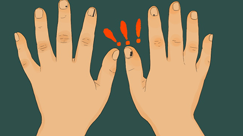
Onychomycosis results from fungal infection. Contributing factors include nail trauma, tinea pedis (athlete’s foot), impaired local or lymphatic circulation, and immune dysfunction. Clinical manifestations vary widely: infected nail plates may become thickened, opaque, separated from the nail bed, atrophic, discolored, detached, irregularly contoured, or associated with paronychia. Symptoms are often mild but may include slight pain or bleeding.
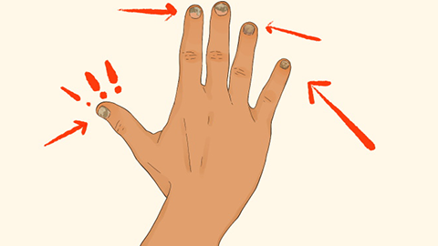
Onychomycosis is classified into four clinical types: distal lateral subungual onychomycosis (DLSO), white superficial onychomycosis (WSO), proximal subungual onychomycosis (PSO), and total dystrophic onychomycosis (TDO)—also termed “nail dystrophy.” In early-stage infection, affected nails typically show localized thickening, discoloration (often yellowing), loss of luster, surface roughness, and uneven contour. As the disease progresses, the affected area expands, eventually evolving over time into total dystrophic onychomycosis. We hope this information proves helpful!

