The Orthodontic Treatment Process
Dec 21, 2021
Source: Cainiu Health

Dr. Qiang Yanli
Introduction
Orthodontic Treatment Process:
Consultation with a Dentist: Development of a personalized treatment plan. Primary options include invisible aligners, ceramic braces, and metal braces—select the most suitable option based on individual needs.
Radiographic Imaging: X-ray imaging of the teeth and skull (including the jawbones). This step aims to classify the type of dentofacial and dental arch deformities and assess skeletal growth patterns, thereby facilitating the formulation of an accurate and effective orthodontic treatment plan.
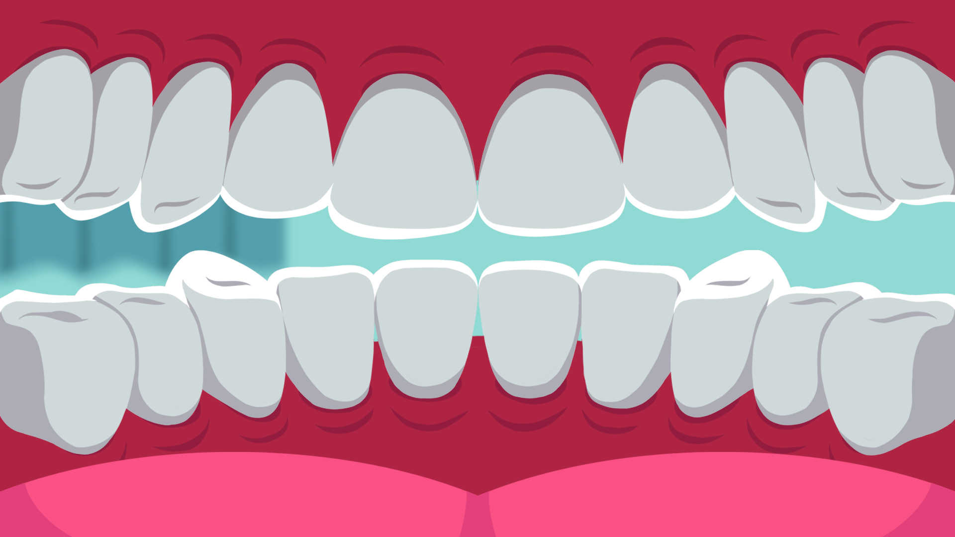
Teeth are a vital part of our body. Dental health not only affects overall physical well-being but also influences facial aesthetics—particularly the alignment of teeth, which significantly impacts one’s appearance. Consequently, dental concerns have drawn increasing public attention. Today, multiple orthodontic treatment options are available for malaligned teeth, and orthodontic correction is suitable for individuals of all ages. As long as patients follow their orthodontist’s instructions carefully throughout treatment, successful outcomes can be achieved at any stage of life.
Orthodontic Treatment Process
Consultation with an Orthodontist
Development of a personalized treatment plan. Common options include clear aligners, ceramic braces, and traditional metal braces. Patients should select the most appropriate option based on their individual needs and preferences.
Radiographic Imaging
X-ray imaging of teeth and craniofacial structures (including panoramic and cephalometric radiographs). These images help determine the type and severity of dental and skeletal malocclusion, assess jaw growth patterns, and guide the formulation of an accurate, evidence-based treatment plan.
Dental Impressions
A fast-setting impression material is placed over the teeth; after biting down for 1–2 minutes, the impression is removed and used to fabricate precise plaster or digital models of the patient’s dentition. These models serve as baseline references for treatment planning and subsequent progress evaluation.
Photographic Documentation
Comprehensive intraoral and extraoral photographs are taken from multiple angles prior to treatment initiation. These images support diagnostic analysis, treatment monitoring, outcome assessment, and medical record documentation.
Tooth Extraction
Based on the treatment plan, certain teeth may be extracted in stages. Most patients require extraction of approximately four premolars. In more complex cases—such as severe crowding or presence of multiple impacted wisdom teeth—up to eight teeth may be removed. Some patients may not require extractions at all; instead, minor interproximal enamel reduction (“stripping”) may suffice. Any existing dental caries will first be restored, and periodontal therapy (e.g., scaling and root planing) will be performed if calculus or gingival inflammation is present. The primary purpose of extractions is to create space for proper alignment of crowded teeth; the resulting gaps will gradually close during active treatment, so patients need not worry about permanent spacing.
Separation of Teeth (Tooth Separation)
Tooth separation may occur concurrently with or following extractions. Elastic separators (spacers) are placed between selected posterior teeth (typically molars and premolars) and worn for approximately one week. This creates slight interdental space to facilitate precise bracket placement. Patients may experience mild soreness, sensitivity, or discomfort when biting down during this phase.
Brace Placement
Once tooth separation is complete, braces are bonded onto the teeth. During the initial adjustment period, mild oral irritation or gum tenderness may occur, especially within the first week. These symptoms typically subside spontaneously. For patients experiencing recurrent aphthous ulcers, topical application of Guilin Watermelon Frost spray before bedtime—along with supplementation of vitamin B complex and zinc—may provide symptomatic relief.
Follow-up Appointments
Routine check-ups are generally scheduled every four weeks. Your orthodontist will confirm specific appointment times. During these visits, adjustments—including wire changes or activation of elastic modules—are performed to ensure controlled, predictable tooth movement according to the treatment plan.
Elastic Band Application
The rate of dental alignment varies depending on individual biological response, complexity of malocclusion, and clinical expertise. Noticeable improvement is often evident within one month, and significant alignment is typically achieved by six months. Following initial alignment, elastics (rubber bands) are introduced to close extraction spaces and refine occlusion. Elastics must be removed during meals and replaced afterward. Their biomechanical effect is highly effective in achieving optimal interarch relationships.
Final Alignment and Stabilization
Space closure coincides with midline correction. Once interdental spaces are fully closed and dental midlines are symmetrically aligned, patients achieve a harmonious, aesthetically pleasing smile. However, braces must remain in place for additional stabilization to prevent relapse—allowing newly positioned teeth and surrounding periodontal tissues time to adapt and mature.
Brace Removal
The most exciting moment has arrived! For optimal results, consider professional teeth cleaning or whitening post-treatment to enhance your radiant, confident smile.
When undergoing orthodontic treatment, it is essential to consult an experienced, board-certified orthodontist. Prior to initiating treatment, a comprehensive oral examination—including clinical assessment, radiographic imaging, and diagnostic records—is mandatory. Based on these findings, the orthodontist will formulate an individualized, scientifically sound treatment plan—ensuring safe, efficient, and esthetically satisfying outcomes.